45 simple microscope diagram with labels
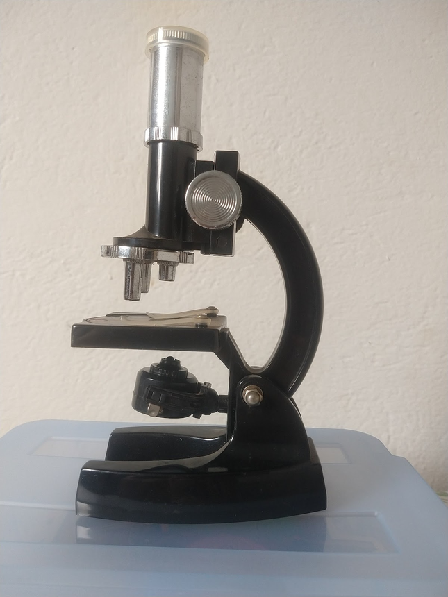
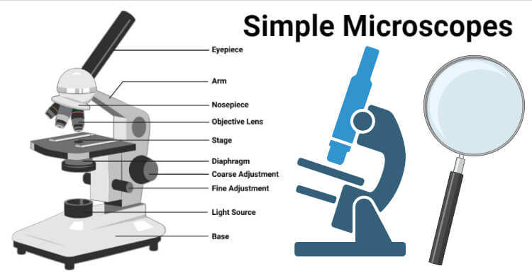
Microscope Types (with labeled diagrams) and Functions Simple microscope labeled diagram Simple microscope functions It is used in industrial applications like: Watchmakers to assemble watches Cloth industry to count the number of threads or fibers in a cloth Jewelers to examine the finer parts of jewelry Miniature artists to examine and build their work Also used to inspect finer details on products Simple epithelium: Location, function, structure | Kenhub Simple epithelium can be divided into 4 major classes, depending on the shapes of constituent cells. The cells found in this epithelium type are flat and thin, making simple squamous epithelium ideal for lining areas where passive diffusion of gases occur.Areas where it can be found include: skin, capillary walls, glomeruli, pericardial lining, pleural lining, peritoneal cavity lining, and ...
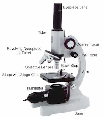
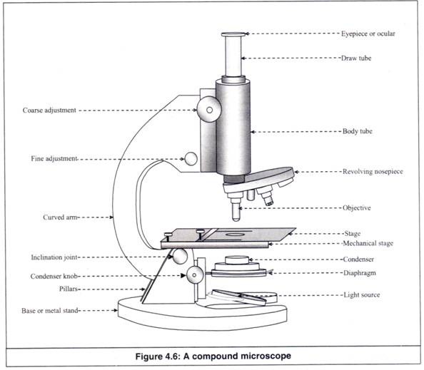
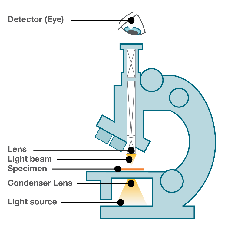
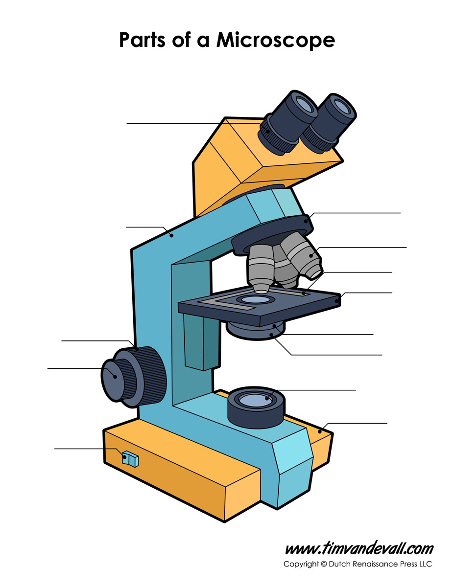
15 Microscope Parts with Diagram, Location and Function - Study Read A compound microscope has about 15 parts that assist in viewing with a naked eye, a sample holder, a magnifying lens, and a light source. For the convenience of study, we can divide them based on their purpose in the instrument like. A) Parts that assist in viewing the object. B) Part that helps in the adjustment of lenses for a clear view.

Simple microscope diagram with labels
Microscope, Microscope Parts, Labeled Diagram, and Functions The Microscopes parts divided into three different structural parts Head, Base, and Arms. Head/Body: It contain the optical parts in the upper part of the microscope. Arm: It supports the tube and connects it to the base. Base: The bottom of the microscope, used for support. Optical Components of Microscope Spectroscope Diagram, Parts, & Function - Study.com Learn the spectroscope definition, what spectroscopes do, and how spectroscopes work. Explore spectroscopes parts and functions and see a spectroscope diagram. Difference between Simple Microscope and Compound Microscope This allows the observers to view the object. The diagram given below shows the structure of a simple microscope with all labeled parts: The formula for the magnification power of a simple microscope is given as follows below: M = 1 + D F Where M is the magnification power of the lens. D is the distance of an object.
Simple microscope diagram with labels. Animal Cell Labeling Quiz Questions And Answers - ProProfs Animal cell labeling can be tricky at first, so why don't we start you off relatively easy with this animal cell part labeling quiz! In this one, we'll be giving you a question referring to a given diagram and asking you to label it. Simple, right? Let's find out how many you get right. All the best! Let's go! Questions and Answers. 1. Parts of the Microscope with Labeling (also Free Printouts) A microscope is one of the invaluable tools in the laboratory setting. It is used to observe things that cannot be seen by the naked eye. Table of Contents 1. Eyepiece 2. Body tube/Head 3. Turret/Nose piece 4. Objective lenses 5. Knobs (fine and coarse) 6. Stage and stage clips 7. Aperture 9. Condenser 10. Condenser focus knob 11. Iris diaphragm label the microscope answers - Alex Becker Marketing This resource contains 1 worksheet for students to label the parts of a microscope and 1 worksheet for students to complete a chart detailing the functions of each microscope part. Answer key included. Click to visit MICROSCOPE PARTS AND FUNCTIONS WORKSHEET.doc - Name: 2. Diaphragm This helps gather light from the mirror and focus it on the stage. Microscope Diagram Worksheet - Stock Walker Write the letter on the line that represents each part of the microscope. Using the terms listed below, label the microscope diagram. When you can identify a part of the microscope place the . There is a printable worksheet available for download here so you can take the . Use the words from this word list to identify the parts of the microscope.
Microscope: Types of Microscope, Parts, Uses, Diagram - Embibe A simple microscope presently consists of a magnifying glass with a double convex lens with a short focal length. Examples of this kind of instrument include the hand lens and reading lens. When an object is kept near the lens, then at its principal focus, an image is produced, which is erect and bigger than the original object. Cells Diagram | Science Illustration Solutions - Edrawsoft Cells Diagram Software Edraw is all-in-one diagram that enables you to draw all kinds of scientific diagrams. It includes many ready-made cells diagram tempates. If you are on short time, you may simply edit the cell diagrams templates with just a few clicks, then you will get your own cells diagram in minutes. Download Cells Diagram Software Testes: Anatomy, definition and diagram | Kenhub Testis. 1/5. The testes (testicles) are male reproductive glands found in a saccular extension of the anterior abdominal wall called the scrotum. They are in ovoid shape, sized four to six centimeters in length. Testes develop retroperitoneally on the posterior abdominal wall and descend to scrotum before birth. Microscope Quiz: How Much You Know About Microscope Parts ... - ProProfs Holds the slide in place. C. It is used to support the microscope when carried. 2. Stage clips: A. Magnification ranges from 10x to 40x. B. Holds the slide in place. C. Moves the stage up and down for focusing. 3. Fine adjustment knob: A. Moves the stage slightly to sharpen the image. B. Holds the high and low power objectives.
Simple Microscope- Definition, Principle, Magnification, Parts ... Optical Parts of Simple Microscope These parts are involved in passing the light through the object (specimen) and magnifying its size. The components of the optical parts are as follows: 1. Mirror A plano-concave mirror is fitted below the stage to the vertical rod using a frame. It focuses the surrounding light on the object to be observed. 2. Simple Microscope - Parts, Functions, Diagram and Labelling Home / Microscopes Simple Microscope - Parts, Functions, Diagram and Labelling By Editorial Team March 7, 2022 A microscope is one of the commonly used equipment in a laboratory setting. A microscope is an optical instrument used to magnify an image of a tiny object; objects that are not visible to the human eyes. Table of Contents Simple cuboidal epithelium- structure, functions, examples Owing to the shape of the cells, the primary functions of the simple cuboidal epithelium are secretion, absorption, and covering. 1. Covering/ Protection. They function as a covering for several organs providing protection against damage and other chemicals. The cuboidal epithelium in the ovaries forms the ovarian surface that functions in ... Pseudostratified Columnar Epithelium under a Microscope with a Labeled ... Pseudostratified Columnar Epithelium under a Microscope with a Labeled Diagram 06/04/2022 by anatomylearner The pseudostratified columnar epithelium comprises a single layer of cells but seems to be multilayered. It is because different cellular heights and nuclei are also placed at a different levels.
Difference between Simple and Compound Microscope ...
Simple Squamous Epithelium under a Microscope with a Labeled Diagram ... Simple squamous epithelium under microscope labeled in renal corpuscle The cortex of a kidney consists of renal corpuscles and the convoluted tubule, straight tubules, nephrons, connecting tubules, and collecting ducts. You will find the medullary ray in the medulla of the kidney that comprises straight tubules and collecting ducts.
Microscope Parts & Specifications | Microscope World Resources
Basic Microscope Diagram - microscope diagram purposegames, images 01 ... Basic Microscope Diagram - 15 images - label the neuron clip art at vector clip art online, microscope diagram fill online printable fillable blank pdffiller, animal anatomy biology4isc, images 01 introduction and terminology basic human anatomy,
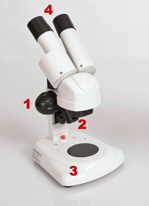
Compound and Stereo- microscopes - Microscopes 4 Schools
Plant Cell: Diagram, Types and Functions - Embibe Exams Observe the labelled diagram of plant cell structure as given below: Are Plant Cells Prokaryotic or Eukaryotic? The cell is the basic structural and functional unit of life in all living organisms. The cells can be divided into two major groups - Prokaryotic and Eukaryotic. The difference between both the cells are explained below:

How to Draw a Simple Microscope Diagram
Simple Cuboidal Epithelium: Location & Function - Study.com Simple Cuboidal Epithelium: Labeled Diagram. Simple cuboidal epithelial cells are shaped like cubes, and the nucleus of each cell is large and located close to the center of the cell. This is ...
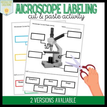
Microscope Labeling Activity
Microscopy- History, Classification, Terms, Diagram - The Biology Notes A simple or compound light microscope is used in this technique. It uses transmitted visible light to develop magnified images. It has a low contrasting capacity, low optical resolution, requires staining and has a limited magnification of around 1300X. It is simple and can be used to observe living cells and microorganisms. 2 Dark Field Microscopy

Compound Microscope Parts, Functions, and Labeled Diagram ...
Simple Microscope Class 12, Definition, Magnification, Working, Parts ... Figure: This is the labeled diagram of a simple microscope showing its different parts source credit: microscopeclub.com Main parts of the simple microscope Eyepiece: This is the lens through which the observer views the image. It is generally positioned at the top. Base: It is the bottom part of the microscope, and holds the structure.

2.2 Molecular make up of cells | Cells: the basic units of ...
Ovary histology slide diagram and identification points Ovary histology slide - identification points. Presence of peripheral cortex showing ovarian follicles at different stages of maturation. There is a centrally placed medulla containing blood vessels. Presence of the primordial follicle containing primary oocyte surrounded by squamous follicular cells. The primary follicle contains a primary ...

Lecture 1: Intro to EM for Biologists | Electron Microscope Unit
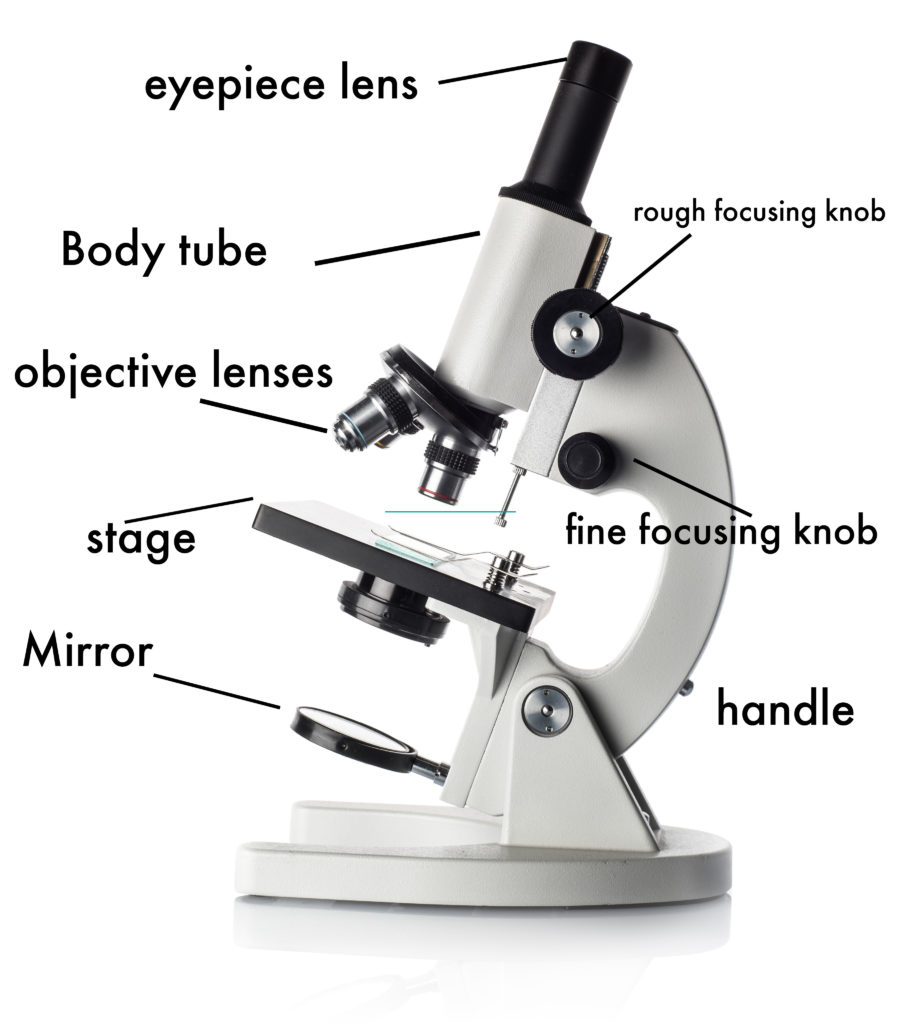
Parts of a microscope with functions and labeled diagram - Microbe Notes Figure: Diagram of parts of a microscope There are three structural parts of the microscope i.e. head, base, and arm. Head - This is also known as the body. It carries the optical parts in the upper part of the microscope. Base - It acts as microscopes support. It also carries microscopic illuminators.

Adebiyi Olusoji Aderemi (adebiyiolusoji) - Profile | Pinterest
Simple Microscope - Diagram (Parts labelled), Principle, Formula and Uses A simple microscope consists of Optical parts Mechanical parts Labeled Diagram of simple microscope parts Optical parts The optical parts of a simple microscope include Lens Mirror Eyepiece Lens A simple microscope uses biconvex lens to magnify the image of a specimen under focus.

Parts of a microscope with functions and labeled diagram
microscope | Types, Parts, History, Diagram, & Facts Optical microscopes can be simple, consisting of a single lens, or compound, consisting of several optical components in line. The hand magnifying glass can magnify about 3 to 20×. Single-lensed simple microscopes can magnify up to 300×—and are capable of revealing bacteria —while compound microscopes can magnify up to 2,000×.

Free Microscope Drawing, Download Free Microscope Drawing png ...
Animal Cells: Labelled Diagram, Definitions, and Structure - Research Tweet Microscope, Microscope Parts, Labeled Diagram, and Functions September 3, 2022 View Details » Pinocytosis: Definition, Types, Features, and Functions June 29, 2022 View Details » ... Simple Diffusion vs Facilitated Diffusion The movement of molecules from the plasma membrane with the help of transporter protein such as carrier is called as ...

Simple Microscope Principle and Working
Difference between Simple Microscope and Compound Microscope This allows the observers to view the object. The diagram given below shows the structure of a simple microscope with all labeled parts: The formula for the magnification power of a simple microscope is given as follows below: M = 1 + D F Where M is the magnification power of the lens. D is the distance of an object.

Compound Microscope Parts – Labeled Diagram and their ...
Spectroscope Diagram, Parts, & Function - Study.com Learn the spectroscope definition, what spectroscopes do, and how spectroscopes work. Explore spectroscopes parts and functions and see a spectroscope diagram.
Free Microscope Drawing, Download Free Microscope Drawing png ...
Microscope, Microscope Parts, Labeled Diagram, and Functions The Microscopes parts divided into three different structural parts Head, Base, and Arms. Head/Body: It contain the optical parts in the upper part of the microscope. Arm: It supports the tube and connects it to the base. Base: The bottom of the microscope, used for support. Optical Components of Microscope

Compound Microscope- Definition, Labeled Diagram, Principle ...

Simple Microscope: Definition, working, diagram, properties, Uses

discovering the simple microscope

The Microscope

Fluorescence microscope - Wikipedia
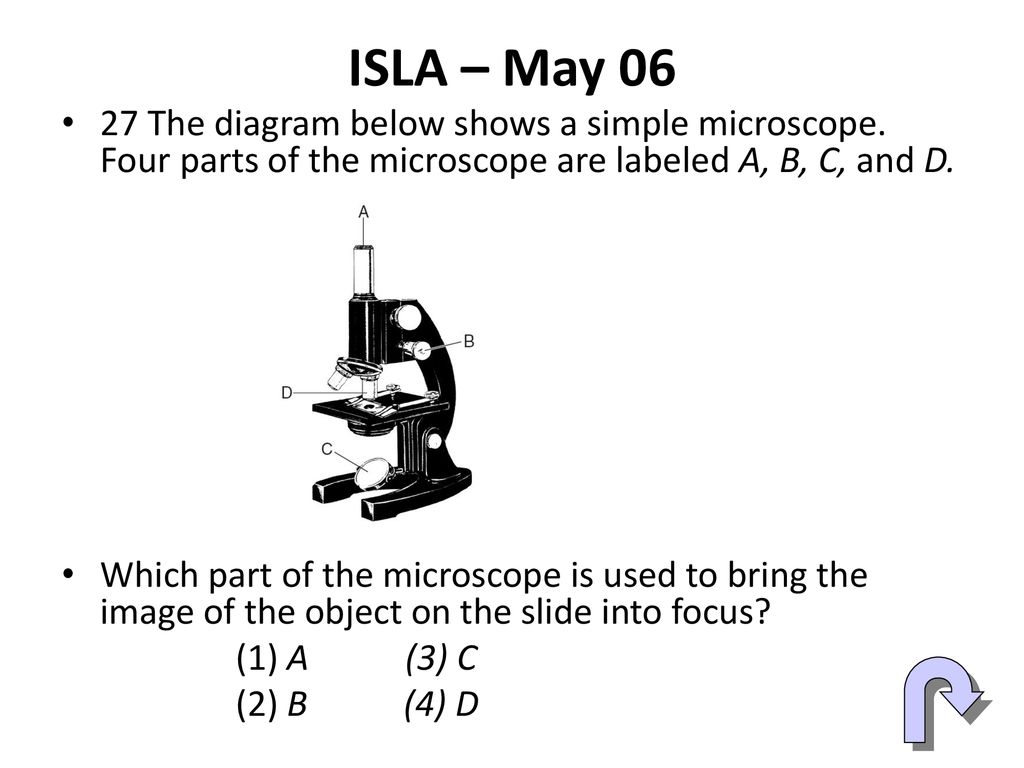
Teaching a Lesson LE Lesson Using Regents Diagrams - ppt download
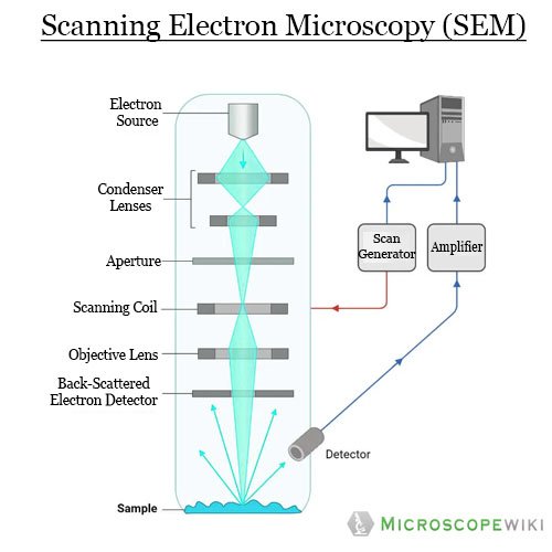
Electron Microscope Principle, Uses, Types and Images ...

Simple Microscope - Parts, Functions, Diagram and Labelling ...

Compound Microscope Parts, Functions, and Labeled Diagram ...

Compound Microscope Parts, Diagram Definition, Application ...

Optical Instruments – Track2Training

How to Choose the Perfect Student Microscope — BioBox Labs
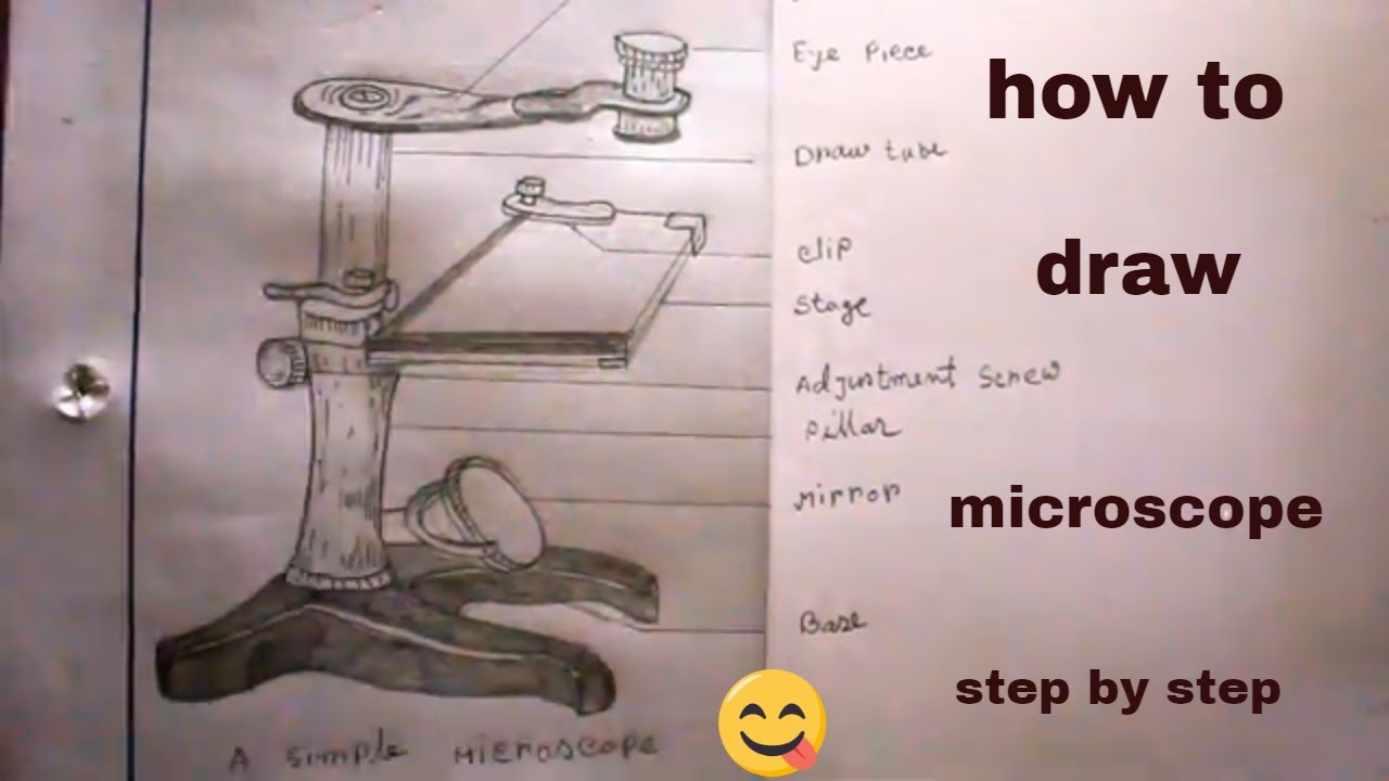
How TO Draw simple microscope step by step/simple microscope drawing/for science project

How to draw Microscope diagram for beginners - step by step

Simple Microscope - Diagram (Parts labelled), Principle ...
How to Use a Microscope
Light Microscope vs Electron Microscope - Life in Atomic ...
Free Microscope Drawing, Download Free Microscope Drawing png ...

Compound and Stereo- microscopes - Microscopes 4 Schools
microscope | Types, Parts, History, Diagram, & Facts | Britannica

Microscope Parts and Functions
Microscopy: Intro to microscopes & how they work (article ...
Simple Microscope - Definition, Diagram, FAQs

The Compound Light Microscope Diagram | Quizlet
microscope - Kids | Britannica Kids | Homework Help
Microscope Parts and Functions

Compound Microscope Parts – Labeled Diagram and their ...
Microscope Diagram Labeled, Unlabeled and Blank | Parts of a ...
Simple Microscope- Definition, Principle, Magnification ...

Parts of Dissecting Microscope | Botany
Molecular Expressions: Microscopy Publications - Fascinating ...
Post a Comment for "45 simple microscope diagram with labels"