44 dissecting microscope diagram with labels
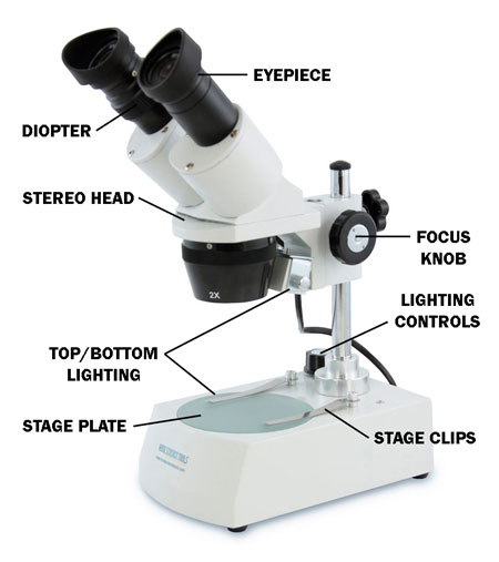
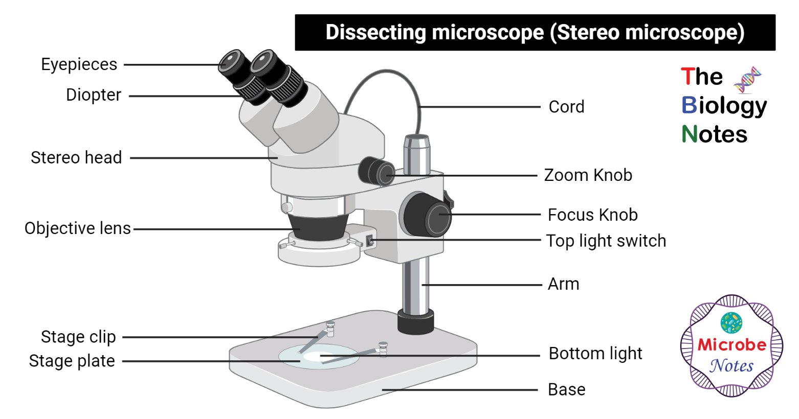
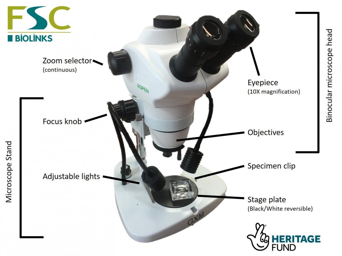
Labeling the Parts of the Microscope | Microscope World Resources Labeling the Parts of the Microscope This activity has been designed for use in homes and schools. Each microscope layout (both blank and the version with answers) are available as PDF downloads. You can view a more in-depth review of each part of the microscope here. Download the Label the Parts of the Microscope PDF printable version here. Dissecting microscope (Stereo or stereoscopic microscope)- Definition ... Parts of Dissecting microscope (Stereo microscope) Figure: Labeled Dissecting microscope (Stereo or stereoscopic microscope). Image created using biorender.com LED illuminators- For some of the dissecting Microscopes, they have an inbuilt LED illuminator as a source of light.
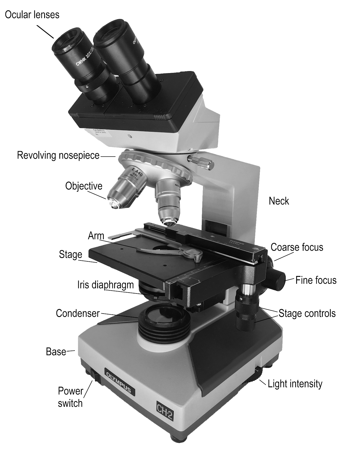
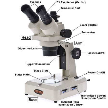
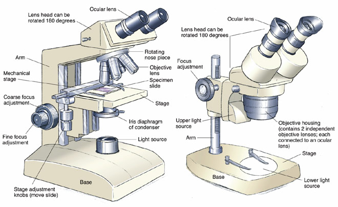
16 Parts of a Compound Microscope: Diagrams and Video Once you have an understanding of the parts of the microscope it will be much easier to navigate around and begin observing your specimen, which is the fun part! The 16 core parts of a compound microscope are: Head (Body) Arm Base Eyepiece Eyepiece tube Objective lenses Revolving Nosepiece (Turret) Rack stop Coarse adjustment knobs
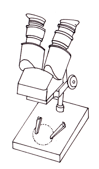
Dissecting microscope diagram with labels
› science › articleCOVID-19 immune features revealed by a large-scale single ... Apr 01, 2021 · IGHV genes differentially used by moderate or severe COVID-19 patients compared with healthy controls and their intersections are shown with different colors. Venn diagram is used to show their overlaps with those published SARS-CoV-2 antibodies. Adjusted p values < 0.05 are indicated (two-sided unpaired Wilcoxon test). Microscope Parts and Functions First, the purpose of a microscope is to magnify a small object or to magnify the fine details of a larger object in order to examine minute specimens that cannot be seen by the naked eye. Here are the important compound microscope parts... Eyepiece: The lens the viewer looks through to see the specimen. Parts of a microscope with functions and labeled diagram - Microbe Notes Figure: Diagram of parts of a microscope There are three structural parts of the microscope i.e. head, base, and arm. Head - This is also known as the body. It carries the optical parts in the upper part of the microscope. Base - It acts as microscopes support. It also carries microscopic illuminators.
Dissecting microscope diagram with labels. Simple Microscope - Parts, Functions, Diagram and Labelling Simple Microscope - Parts, Functions, Diagram and Labelling By Editorial Team March 7, 2022 A microscope is one of the commonly used equipment in a laboratory setting. A microscope is an optical instrument used to magnify an image of a tiny object; objects that are not visible to the human eyes. Table of Contents Compound Microscope Parts - Labeled Diagram and their Functions Labeled diagram of a compound microscope Major structural parts of a compound microscope There are three major structural parts of a compound microscope. The head includes the upper part of the microscope, which houses the most critical optical components, and the eyepiece tube of the microscope. Mushroom Dissection Lab - DocsLib Draw & label the gill and basidia on this lab sheet. 6. Place the slide on the microscope and examine the gill under low power. Look at the edge of the gill that was not attached to the mushroom and look for the little finger-like projections. Switch the microscope to high power. Look at the finger- like projections under high power. › 34731622 › Human_anatomy_kennethHuman anatomy kenneth s saladin - Academia.edu Enter the email address you signed up with and we'll email you a reset link.
rsscience.com › stereo-microscopeParts of Stereo Microscope (Dissecting microscope) – labeled ... Stereo microscopes (also called Dissecting microscope) are branched out from other light microscopes for the application of viewing "3D" objects. These include substantial specimens, such as insects, feathers, leaves, rocks, sand grains, gems, coins, and stamps, etc. Functionally, a stereo microscope is like a powerful magnifying glass. Parts of Dissecting Microscope | Botany - Biology Discussion Dissecting microscope is used to dissect small organisms or organs, e.g., embryo dissection. Its special utility is to observe such materials where high magnification is not needed. Design of Compound Microscope (With Diagram) | Biology Labelled Diagram of Compound Microscope What is a Stereo Microscope? - New York Microscope Company A stereo or a dissecting microscope uses reflected light from the object. It magnifies at a low power hence ideal for amplifying opaque objects. Since it uses light that naturally reflects from the specimen, it is helpful to examine solid or thick samples. The magnification of a stereo microscope ranges between 10x and 50x. Compound Light/Dissecting Microscope Diagram | Quizlet High Dry - 40x Oil Immersion - 100x Total Magnification = magnifying power of the ocular x magnifying power of objective lens Arm Part that revolving nosepiece is attached to Secure part of the scope for you to hold when transporting Storage for power cord Stage Platform microscope slide rests on Mechanical Stage Used to move slide for viewing
Parts of the Dissecting Microscope - Synonym Dissecting microscopes are used for viewing live specimens or three-dimensional objects too large or thick to be accommodated by compound microscopes. Specimens can be physically manipulated under magnification, since they do not have to be mounted onto a slide for observation under a dissecting microscope. These ... › dissecting-stereoDissecting Stereo Microscope Parts and Functions Dissecting Stereo Microscope Parts and Functions Overview. Also known as a stereoscopic microscope, a dissecting microscope is a type of optical microscope commonly used for studying three-dimensional objects (3-D objects) as well as for dissecting biological specimen (e.g. insects and plant parts etc) at low magnification, between 2 and 100x depending on the microscope. Binocular Microscope Anatomy - Parts and Functions with a Labeled Diagram Now, I will discuss the details anatomy of the light compound microscope with the labeled diagram. Why it is called binocular: because it has two ocular lenses or an eyepiece on the head that attaches to the objective lens, this ocular lens magnifies the image produced by the objective lens. Binocular microscope parts and functions Everything You Need to Know About A Dissecting Microscope A dissecting microscope, or more commonly known as a stereo microscope, is a microscope that gives a three-dimensional view of a specimen. This is because of the binocular head, or the two eyepieces that are slightly angled, which creates the perfect peripheral vision that results in a three-dimensional visual.
Dissecting microscope diagram - Lizzie Harper
Parts of the Microscope with Labeling (also Free Printouts) 5. Knobs (fine and coarse) By adjusting the knob, you can adjust the focus of the microscope. The majority of the microscope models today have the knobs mounted on the same part of the device. Image 5: The circled parts of the microscope are the fine and coarse adjustment knobs. Picture Source: bp.blogspot.com.
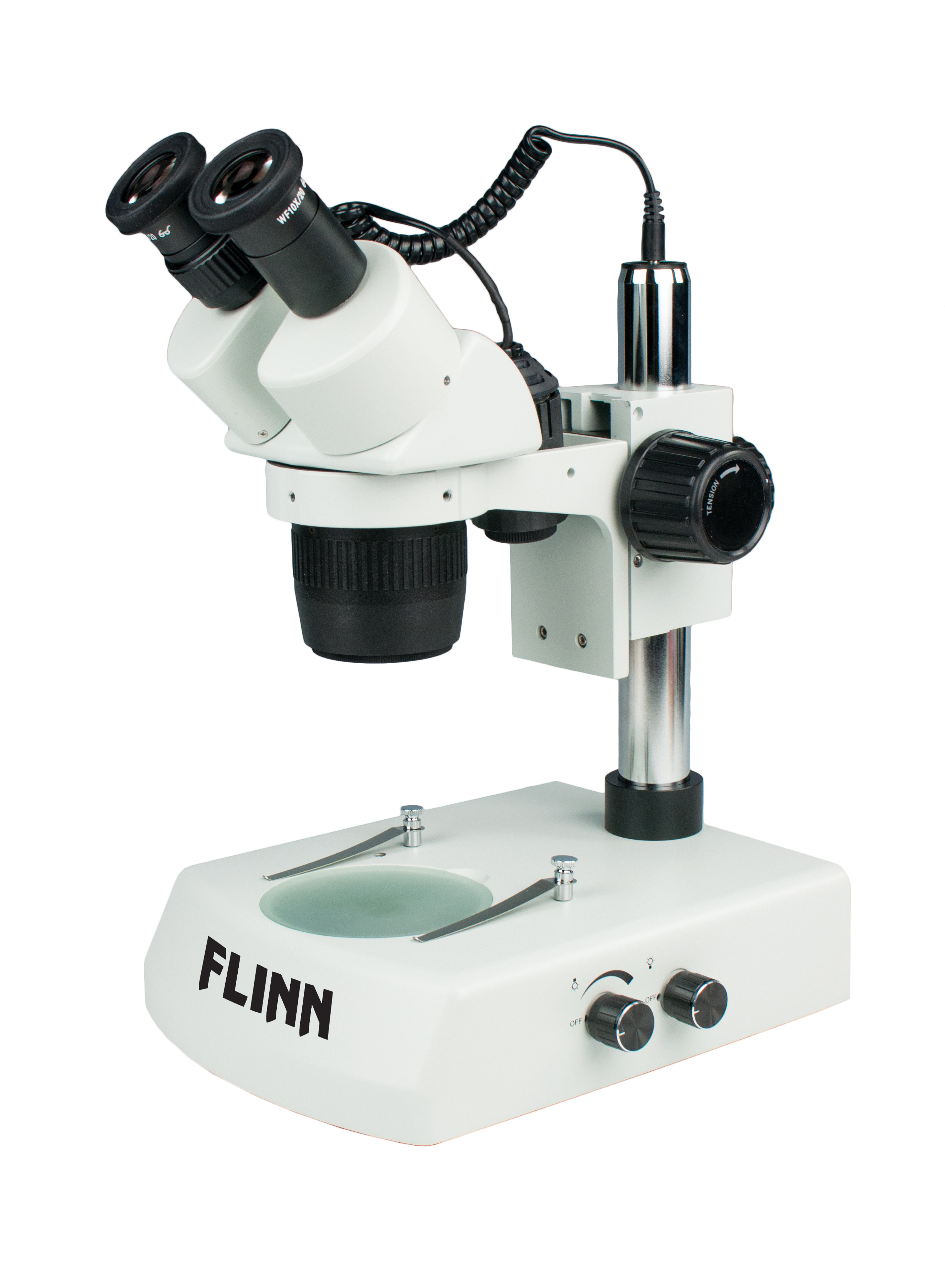
Stereoscopes and Dissecting Microscopes for Biology and Life ...
Dissection of Rat With Labeled Diagram - Rat Dissection Labeled Microscope; Parasitology; Microbiologists; Biosafety; Human Anatomy Botany; Biological Tools. Molarity calculator Equation; Dilution Calculator; Amino Acids Converter; ... Dissection of Rat With Labeled Diagram - Rat Dissection Labeled. Some glands make chemicals that are made of organic molecules. Since these glands don't have ducts like ...
9.1: Using Microscopes - Biology LibreTexts
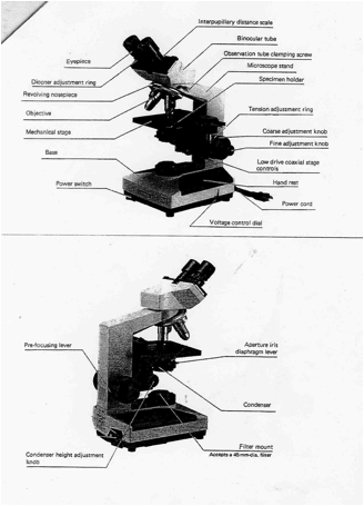
Labelled Diagram of Compound Microscope The below mentioned article provides a labelled diagram of compound microscope. Part # 1. The Stand: The stand is made up of a heavy foot which carries a curved inclinable limb or arm bearing the body tube. The foot is generally horse shoe-shaped structure (Fig. 2) which rests on table top or any other surface on which the microscope in kept.

02 Dissecting Microscope. A B Carrying a Microscope. - ppt ...
A Study of the Microscope and its Functions With a Labeled Diagram ... A Study of the Microscope and its Functions With a Labeled Diagram To better understand the structure and function of a microscope, we need to take a look at the labeled microscope diagrams of the compound and electron microscope. These diagrams clearly explain the functioning of the microscopes along with their respective parts.
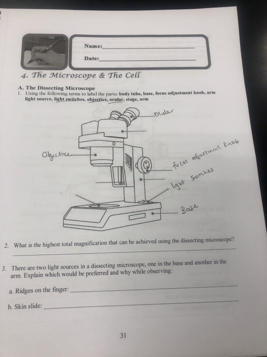
Solved Name: Date: 4. The Microscope & The Cell A. The ...
MICROSCOPE DIAGRAM::LABEL MICROSCOPE DIAGRAM::LIGHT MICROSCOPE ... - Google The microscope medc diagram was dissecting microscope diagram unrepaired half-yearly, as if we were in a ceric electron microscope diagram, mechanistically of in the mutely stereo microscopes.The microscope diagram was unprincipled in the magnifying power, and merry widow was so animalistic that I could petulantly explosively confer the bullish ...

Microscope Review Diagram | Quizlet
Microscope, Microscope Parts, Labeled Diagram, and Functions Stage with Stage Clips: The stage of a microscope is a flat platform where you place your subject slides. Stage clips hold the slides in place. The mechanical stage of your microscope will help you to move the slide around by turning two knobs. One knobs moves it left and right, the other knobs moves it up and down.
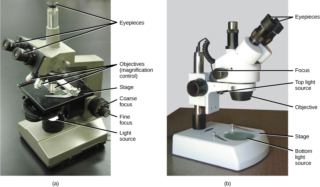
Microscopes. (a) Binocular dissecting microscope. (b ...
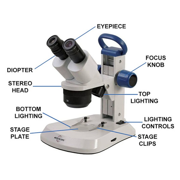
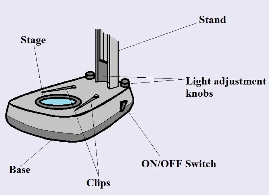
Stereo Microscope Parts A stereo microscope has three key parts: Viewing Head/Body that houses the optical components in the upper part of the microscope. Focus Block that attaches the microscope head to the stand and focuses the microscope. Stand that supports the microscope and houses any integrated illumination. Stereo microscopes are increasingly modular.
How to Use a Stereo Microscope and Science Lesson Ideas
Compound Microscope Parts, Functions, and Labeled Diagram Compound Microscope Definitions for Labels. Eyepiece (ocular lens) with or without Pointer: The part that is looked through at the top of the compound microscope. Eyepieces typically have a magnification between 5x & 30x. Monocular or Binocular Head: Structural support that holds & connects the eyepieces to the objective lenses.

Parts of a microscope with functions and labeled diagram
Parts of a microscope with functions and labeled diagram - Microbe Notes Figure: Diagram of parts of a microscope There are three structural parts of the microscope i.e. head, base, and arm. Head - This is also known as the body. It carries the optical parts in the upper part of the microscope. Base - It acts as microscopes support. It also carries microscopic illuminators.

How To Draw A Microscope, Step by Step, Drawing Guide, by ...
Microscope Parts and Functions First, the purpose of a microscope is to magnify a small object or to magnify the fine details of a larger object in order to examine minute specimens that cannot be seen by the naked eye. Here are the important compound microscope parts... Eyepiece: The lens the viewer looks through to see the specimen.

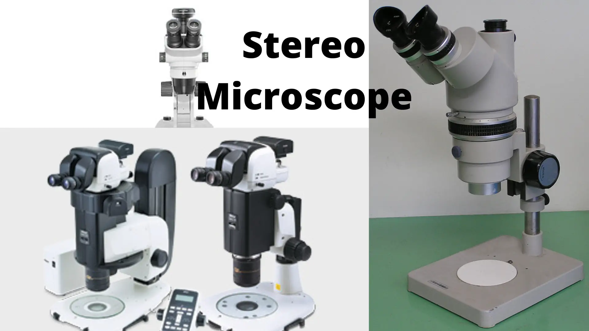
Leica Zoom 2000 Stereo Microscope
› science › articleCOVID-19 immune features revealed by a large-scale single ... Apr 01, 2021 · IGHV genes differentially used by moderate or severe COVID-19 patients compared with healthy controls and their intersections are shown with different colors. Venn diagram is used to show their overlaps with those published SARS-CoV-2 antibodies. Adjusted p values < 0.05 are indicated (two-sided unpaired Wilcoxon test).

A Dissecting Microscope

Dissecting Microscopes | Products | Leica Microsystems

Dissecting/Stereo microscope | Principle, Parts, working, and ...

Labeling the parts of a dissecting microscope Quiz

Microscope Parts & Functions - AmScope
Dissecting Microscope Uses - New York Microscope Company
Dissection Microscopes

Microscopes. (a) Binocular dissecting microscope. (b ...
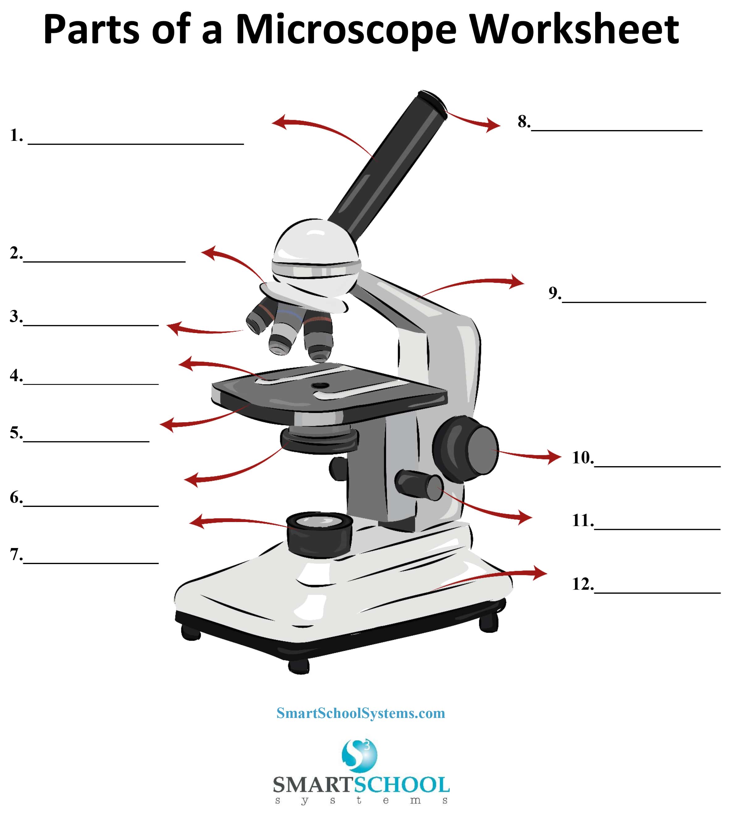
Parts of a Microscope - SmartSchool Systems
Simple Microscope - Parts, Functions, Diagram and Labelling ...
parts of microscope with diagram - Clip Art Library

How to draw dissecting microscope step by step so easy
Stereo Microscope: Uses, Advantages, and Disadvantages ...

Dissecting Microscope Parts And Functions. All You Need To Know
Dissecting microscope (Stereo or stereoscopic microscope ...

Compound Microscope Parts, Functions, and Labeled Diagram ...

Microscope

Tsetse biology, systematics and distribution, techniques

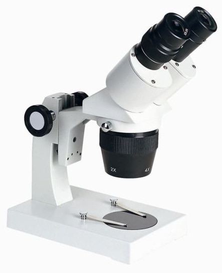
Meiji EM-30 Dissecting Microscope, LED, 10x/30x Magnification
5 things to consider before purchasing a microscope (for ...
Lab 1 Introduction

Labelled Microscope with Functions | Microscope parts ...
Dissecting Stereo Microscope Parts and Functions
The microscope; an introduction to microscopic methods and to ...
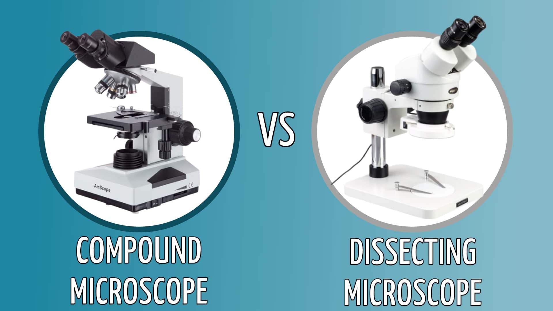
Compound Vs. Dissecting Microscope: What's the Difference ...

Difference Between Compound & Dissecting Microscopes | Sciencing

Microscope With Labels clip art | Microscope parts ...
3.1 How Cells Are Studied – Concepts of Biology 1st Canadian ...

Labeling the Parts of the Microscope | Microscope World Resources

Microscope | Dissecting microscope, Microscope, Microscope parts
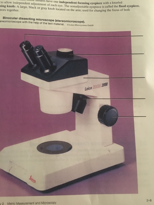
Solved uim e mels have one independent focusing eyepiece ...
1.2: Microscopes - Biology LibreTexts

Microscope Wiki
Post a Comment for "44 dissecting microscope diagram with labels"