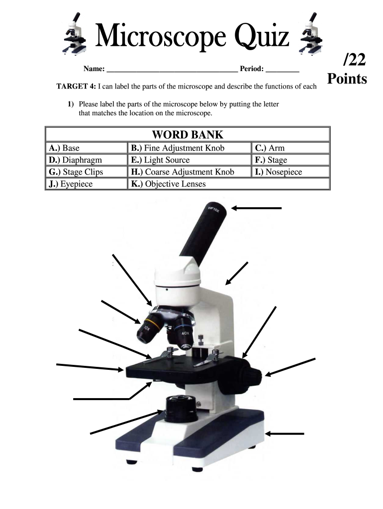
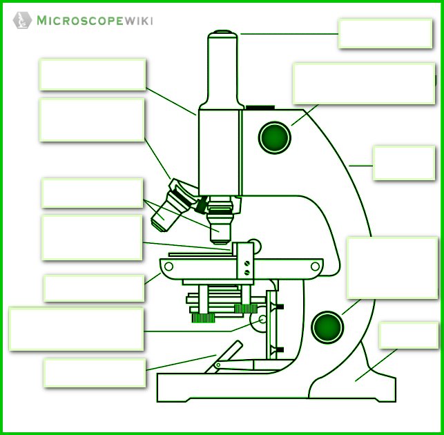
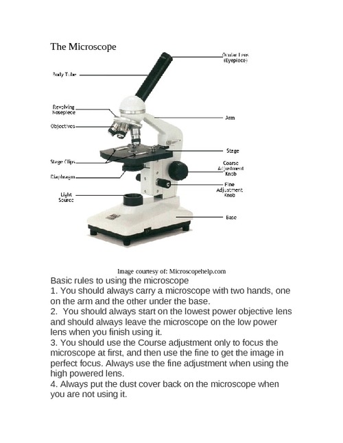
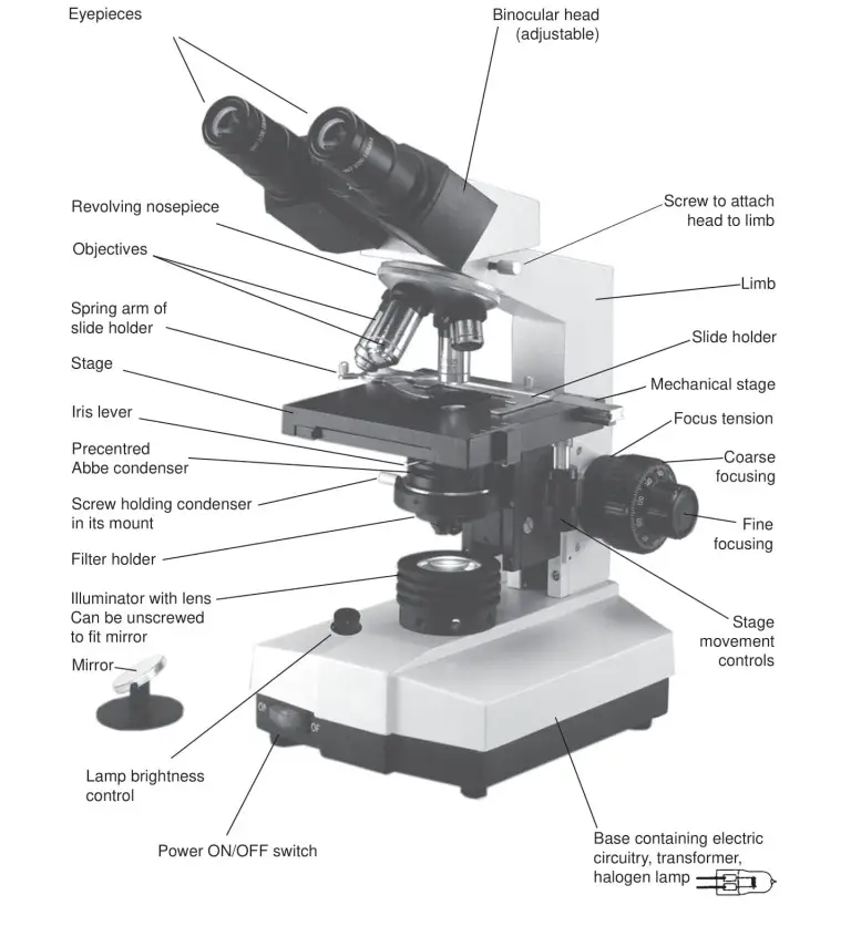
42 labels of a microscope and functions
Light Microscope (Theory) - Amrita Vishwa Vidyapeetham Parts of a Microscope It consists of mainly three parts: Mechanical part - base, c-shaped arm and stage. Magnifying part - objective lens and ocular lens. Illuminating part - sub stage condenser, iris diaphragm, light source. Mechanical part Base: It helps in holding the various parts of microscope. It also contains the light source. Autoclave: Principle, Procedure, Types, Uses - Microbe Online The use of moist heat facilitates the killing of all microorganisms, including heat-resistant endospores which is achieved by heating the materials inside the device at temperatures above the boiling point of water. According to the principle of gas laws, this can be achieved by raising the pressure inside the device.
RBC Blood Test: Normal Ranges and Diagnostic Uses - Verywell Health The red blood cell (RBC) count is a test that measures the number of oxygen-carrying blood cells in your blood. An abnormal RBC test result is often the first sign of an illness. At other times, the test can point the doctor in the direction of a diagnosis if there are symptoms like unexplained fatigue or shortness of breath.

Labels of a microscope and functions
EdU Assay / EdU Staining Proliferation Kit (iFluor 488) (ab219801 ... Dot plot of EdU-488 staining (Y-axis, 488) vs FSC. 10 6 HeLa (Human epithelial cell line from cervix adenocarcinoma) cells were incubated with the stated concentrations of EdU for 3 hours. Control cells (next image) were incubated with media only. Images were acquired on an Accuri C6 Cytometer (BD Biosciences) with cells excited using a 488 nm laser and data analyzed using FlowJo (v10). Motility Test (Theory) - Amrita Vishwa Vidyapeetham Virtual Lab Three methods are employed for motility determination depending on the pathogenic capability of the organisms. For nonpathogens, there are two slide techniques that one might use. For pathogens, tube method can be used. I) Slide methods for non-pathogens include 1. Wet Mount slide 2. Hanging Drop slide 1. Wet Mount slide Corning ® 96 Well Black Polystyrene Microplate - Sigma-Aldrich Corning® | flat bottom clear | black polystyrene plate (Ideal for fluorescent assays) | Tissue Culture (TC)-treated surface | individually wrapped) | sterile | lid
Labels of a microscope and functions. General Structure and Functions of Red Blood Cells 20 to 30 trillion red blood cells ( erythrocytes; RBCs) circulate in the bloodstream of an average adult. (The ring-shaped fat-filled cells in the illustration are called Adipocytes) Red blood cells circulating in the blood stream. RBCs are small, disc-shaped cells that measure 7 - 8 micrometers (μm) in diameter. The diameter of a red blood cell. 1 Form 2 Biology End Term 2 Exams Plus Marking Schemes Interrelationships between organism and their environment. Heredity and variations. The structure below was observed under the light microscope. Identify the cell structure. (1mk) Name the parts labeled A and B (2mks) State two functions of the above structure. (2mks) The diagram below shows a certain cell in living organisms. Self-Taught AI May Have a Lot in Common With the Human Brain Such "supervised" training requires data laboriously labeled by humans, and the neural networks often take shortcuts, learning to associate the labels with minimal and sometimes superficial... › createJoin LiveJournal Password requirements: 6 to 30 characters long; ASCII characters only (characters found on a standard US keyboard); must contain at least 4 different symbols;
› microscopy › enZEISS Axio Observer for Life Science Research Expect a remarkable increase in efficiency with the Axio Observer automation functions. Fast switchable light sources and filters give you highest spectral flexibility and speed. Select the ideal camera to always get the image quality and speed your applications require. The 25 Best STEM Toys of 2022 - The Spruce Every budding scientist needs a microscope, and this one is designed especially for kids ages 3 and up. It magnifies items up to 8x and has a variety of kid-friendly features, such as a chunky focusing knob and extra-large eyepieces for inspecting flowers, bugs, or anything else you can find in your own backyard. Price at time of publish: $18 en.wikipedia.org › wiki › Spinal_cord_injurySpinal cord injury - Wikipedia Human bone marrow derived mesenchymal stem cells seen under phase contrast microscope (63 x magnification) Stem cell transplantation is an important avenue for SCI research: the goal is to replace lost spinal cord cells, allow reconnection in broken neural circuits by regrowing axons, and to create an environment in the tissues that is ... What are spermatogonia and what types are there? - science - 2022 Spermatogenesis is defined as the process by which spermatogonia cells form spermatozoa and, at least in adult mammals, it is a process that occurs continuously until death. It occurs in the testes, initially in structures called seminiferous tubules, which comprise about 90% of the testicular tissue. It has a mitotic and a meiotic phase.
med.libretexts.org › Bookshelves › Anatomy_and4.1: Cell Structure and Function - Medicine LibreTexts Aug 13, 2020 · A cell is the smallest living thing in the human organism, and all living structures in the human body are made of cells. There are hundreds of different types of cells in the human body, which vary in shape (e.g. round, flat, long and thin, short and thick) and size (e.g. small granule cells of the cerebellum in the brain (4 micrometers), up to the huge oocytes (eggs) produced in the female ... X-ray crystallography - Wikipedia The method also revealed the structure and function of many biological molecules, including vitamins, drugs, proteins and nucleic acids such as DNA. X-ray crystallography is still the primary method for characterizing the atomic structure of new materials and in discerning materials that appear similar by other experiments. Smoker's Lung Pictures: Smokers' Lungs vs. Healthy Lungs - MedicineNet A thin membrane called the pleura covers the outer surface of the lung. The air we breathe gets into the lung through an airway (path for air). Figure 1 is a diagram showing the main parts of the airway and lung. The airway consists of the oral and nasal cavities, which connect to the voice box (larynx), which connects to the windpipe (trachea). Cerebellum and brainstem: Anatomy and functions | Kenhub Anatomy of the brain (sagittal view) The cerebellum and brainstem are a testament to the fact that good things do come in small packages, so this article is an overview of their anatomy.Occupying only a fraction of the volume of the cerebrum, these structures are responsible for simplifying every second of your life and keeping you alive.Thanks to them, you can subconsciously and automatically ...

Microscope Components - Science Quiz
Spinal Cord Cross Section | New Health Advisor This makes it possible for the different parts of the CNS communicate with each other. Every bundle of axons is a tract and it transmits specific information. The tracks that move upward are responsible for signals to the brain and the descending tracts send the signals from the brain to neurons throughout the body. 2. Gray Matter

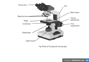
Compound Microscope Parts – Labeled Diagram and their ...
Communicable Diseases Module: 1. Basic Concepts in the Transmission of ... 1.1 What are communicable diseases? As described in the introduction, the organisms that cause communicable diseases are called infectious agents, and their transmission to new uninfected people is what causes communicable diseases; (note that infectious diseases is an interchangeable term). Familiar examples of communicable diseases are malaria and tuberculosis.

Compound Microscope- Definition, Labeled Diagram, Principle ...
N-STORM | Super-Resolution Microscopes | Nikon Microscope Products ... The 3D-Stack function allows multiple 3D STORM images from different Z positions to be captured and stitched into one image to create thicker STORM images. Tubulin of BSC-1 cell labeled with Alexa Fluor ® 647 Tenfold improvement of lateral resolution up to 20nm
Microscope Fill In The Blank - Fill Online, Printable ...
X-ray Powder Diffraction (XRD) - Techniques X-ray powder diffraction (XRD) is a rapid analytical technique primarily used for phase identification of a crystalline material and can provide information on unit cell dimensions. The analyzed material is finely ground, homogenized, and average bulk composition is determined. Fundamental Principles of X-ray Powder Diffraction (XRD)
Types of Microscopes | Microscope World Blog
5 White Blood Cells Types and Their Functions - New Health Advisor Function: As macrophages, monocytes do the job of phagocytosis (cell-eating) of any type of dead cell in the body, whether it is a somatic cell or a dead neutrophil. Because of their large size, they have the ability to digest large foreign particles in a wound unlike other kinds of white blood cells. 4. Eosinophils
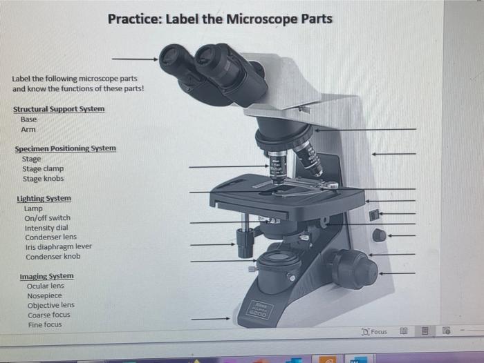
Solved Practice: Label the Microscope Parts Label the | Chegg.com
11 Different Types of Microscopes (With Pictures) - Optics Mag The 11 Types of Microscopes: 1. Light Microscopes The most common type of microscope you're likely to come across, these microscopes rely on lenses and light to illuminate a specimen for optimal image-gathering. They can be used for viewing living cells, insects, for performing dissections, or for clinical blood and tissue assessment. 2.
Monday 10/19/15 AIM: how do the parts of the compound light ...
NIH VideoCasting - Upcoming Events - National Institutes of Health Tuesday, October 11, 2022. 3:00:00 PM - 4:00:00 PM. Ethical & Regulatory Aspects of Clinical Research Session 3: Subject Selection/Recruitment and Retention/Inclusion of Children. Read more. Wednesday, October 12, 2022. 8:30:00 AM - 11:30:00 AM. NIH Combined Federal Campaign (CFC) Kickoff - October 12, 2022 NIH Only.

label the parts of the compound microscope - Brainly.ph
Parts of a Microscope with Their Functions - Microbe Online (40 X): It is a high-power lens. 40X objective lens is used for examination of wet preparations e.g, hanging drop, and ova and cyst examination in the stool. (100 X): It is the oil-immersion lens. The lenses on which oil is used are called oil-immersion lenses.

Labeling the Parts of the Microscope | Microscope activity ...
afn.netAmerican Family News Aug 02, 2022 · American Family News (formerly One News Now) offers news on current events from an evangelical Christian perspective. Our experienced journalists want to glorify God in what we do.

13 parts of the Compound Light Microscope Diagram | Quizlet
› NATIONAL-GEOGRAPHIC-Dual-StudentNational Geographic Dual LED Student Microscope Aug 07, 2017 · National Geographic Dual Microscope has surprisingly clear lens for an inexpensive microscope. The microscope has 10x and 25x lens that are easy to exchange. You simply pull to remove the lens and the next lens slides in. The distance between the two heads is adjustable, but this does not have a Diopter adjustment.

Parts of a Compound Microscope and Their Functions
rsscience.com › stereo-microscopeParts of Stereo Microscope (Dissecting microscope) – labeled ... Unlike a compound microscope that offers a flat image, stereo microscopes give the viewer a 3-dimensional image that you can see the texture of a larger specimen. [In this image] Examples of Stereo & Dissecting microscopes. Major microscope brands (Zeiss, Olympus, Nikon, Amscope, Omano, Leica …) all produce stereomicroscopes.

Parts of a Microscope Labeling Activity
Online Labs for schools - Developed by Amrita Vishwa Vidyapeetham and ... The development of OLabs includes the study and use of mathematical techniques to demonstrate the various complex functions in diverse areas of science. The labs make use of cutting edge simulation technology to create real world lab environments. Thorough study and research is done by research personnel for better understanding of the ...

Parts of a microscope with functions and labeled diagram
DNA - Wikipedia DNA is a long polymer made from repeating units called nucleotides, each of which is usually symbolized by a single letter: either A, T, C, or G. The structure of DNA is dynamic along its length, being capable of coiling into tight loops and other shapes. In all species it is composed of two helical chains, bound to each other by hydrogen bonds. ...
Solved] Associate the functions of the compound light ...
Centriole - Genome.gov Centrioles play a role in organizing microtubules that serve as the cell's skeletal system. They help determine the locations of the nucleus and other organelles within the cell. Narration 00:00 01:25 A centriole is a barrel-shaped organelle which lives normally within the centrosome. The centrosome is the area of the cytoplasm.

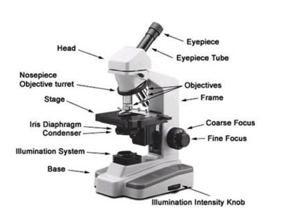
A diagram showing all of the parts of a compound light ...
Plasma Membrane (Cell Membrane) - Genome.gov The plasma membrane, also called the cell membrane, is the membrane found in all cells that separates the interior of the cell from the outside environment. In bacterial and plant cells, a cell wall is attached to the plasma membrane on its outside surface. The plasma membrane consists of a lipid bilayer that is semipermeable.

Parts of the Microscope and Their Uses
3D Laser Scanning Microscope, VK-X200 - KEYENCE Combines the capabilities of an optical microscope, scanning electron microscope and roughness gauge into a single system. Provides non-contact profile, roughness and thickness measurements on nearly any material. Download the catalog for more information: VK-X Series 3D Laser Scanning Confocal Microscope Catalog [PDF:5.88MB] All (132) Support (40)
Simple Microscope - Diagram (Parts labelled), Principle ...
Development of a Technique to Detect Amyloid B-binding Exosomes in a ... The technology developed enables highly sensitive detection of exosomes that retain specific surface molecules from a small amount of blood without the need to learn special techniques, as long as a detection chip and a general-purpose fluorescence microscope are available.

Parts of a Microscope - SmartSchool Systems
Paramedics World | Medical & Paramedical Study Notes ANTIBODIES - DEFINITION, STRUCTURE, FUNCTIONS & CLASSIFICATION. February 27, 2018. SAHIL BATRA. Anatomy & Physiology. ANATOMY AND PHYSIOLOGY NOTES. INTRODUCTION & BASICS OF ANATOMY & PHYSIOLOGY. November 5, 2018. SAHIL BATRA. ANATOMY AND PHYSIOLOGY NOTES. ... What are white label CBD products and what are their benefits? September 30, 2022 ...

16 Basic Parts of Microscope, Function, Names & Labeled Diagram
Corning ® 96 Well Black Polystyrene Microplate - Sigma-Aldrich Corning® | flat bottom clear | black polystyrene plate (Ideal for fluorescent assays) | Tissue Culture (TC)-treated surface | individually wrapped) | sterile | lid
The Microscope Parts and Use
Motility Test (Theory) - Amrita Vishwa Vidyapeetham Virtual Lab Three methods are employed for motility determination depending on the pathogenic capability of the organisms. For nonpathogens, there are two slide techniques that one might use. For pathogens, tube method can be used. I) Slide methods for non-pathogens include 1. Wet Mount slide 2. Hanging Drop slide 1. Wet Mount slide

Compound Microscope Parts, Functions, and Labeled Diagram ...
EdU Assay / EdU Staining Proliferation Kit (iFluor 488) (ab219801 ... Dot plot of EdU-488 staining (Y-axis, 488) vs FSC. 10 6 HeLa (Human epithelial cell line from cervix adenocarcinoma) cells were incubated with the stated concentrations of EdU for 3 hours. Control cells (next image) were incubated with media only. Images were acquired on an Accuri C6 Cytometer (BD Biosciences) with cells excited using a 488 nm laser and data analyzed using FlowJo (v10).

Microscope Parts and Functions
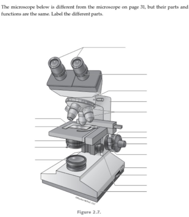
Solved The microscope below is different from the microscope ...

This is a common compound microscope. Label its parts from A ...

What is a Compound Microscope? | Microscope World Blog
Microscope

This is a common compound microscope. What the labelling D ...
Label microscope - Teaching resources
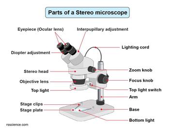
Parts of Stereo Microscope (Dissecting microscope) – labeled ...

Compound Microscope Parts, Functions, and Labeled Diagram ...

Label Microscope Diagram - EnchantedLearning.com

Parts of a microscope with functions and labeled diagram
Parts of a Microscope with Their Functions – Microbe Online

Simple Microscope - Diagram (Parts labelled), Principle ...
Free Microscope Drawing, Download Free Microscope Drawing png ...

Multiple Choice Quiz on Compound Microscope Parts and Functions

List: Parts of a Microscope and their Function | Pathwooded

Parts of a microscope with functions and labeled diagram

label microscope diagram | Charts | Microscope, Anatomy bones ...

Microscope labeling
Parts of a Microscope with Their Functions – Microbe Online

Compound Microscope Parts
Post a Comment for "42 labels of a microscope and functions"