44 external structure of the heart with labels
Human Heart - Anatomy, Functions and Facts about Heart - BYJUS The external structure of the heart has many blood vessels that form a network, with other major vessels emerging from within the structure. The blood vessels typically comprise the following: Veins supply deoxygenated blood to the heart via inferior and superior vena cava, and it eventually drains into the right atrium. External Heart Anatomy labeled.jpg - B. External Anatomy of... View External Heart Anatomy labeled.jpg from BIOL 186 at Messiah. B. External Anatomy of the heart (label diagram, Figure 17.5, p636-637) blue - path of oxygen poor blood arteries away from red- path
Heart anatomy: Structure, valves, coronary vessels | Kenhub The heart is shaped as a quadrangular pyramid, and orientated as if the pyramid has fallen onto one of its sides so that its base faces the posterior thoracic wall, and its apex is pointed toward the anterior thoracic wall.
External structure of the heart with labels
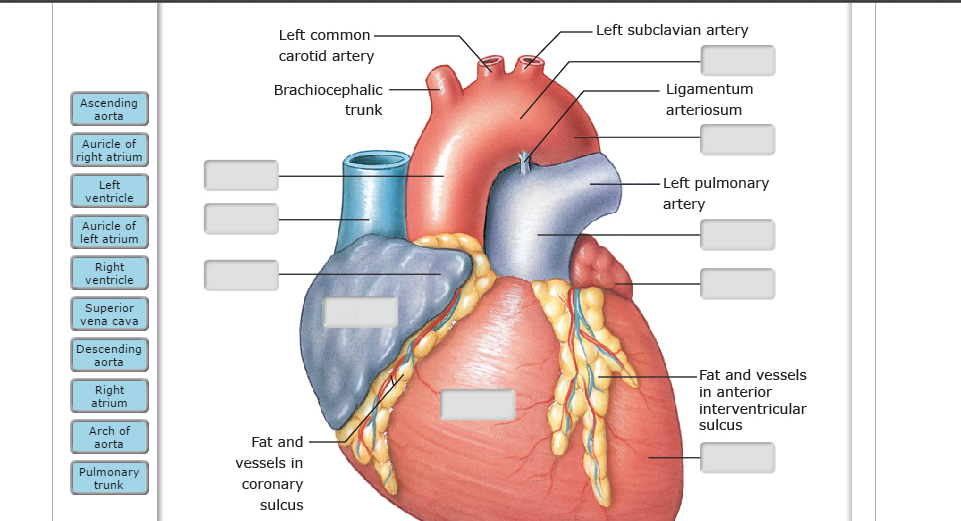
Ch. 19 Circulatory System- heart Flashcards | Quizlet Correctly label the external anatomy of the anterior heart. Place the labels in order denoting the flow of blood through the pulmonary circuit beginning with the right atrium and ending in the left atrioventricular valve. The first and last structures are given. Right atrium 1. tricuspid valve 2. right ventricle 3. pulmonary valve VP Online - Online Drawing Tool - Visual Paradigm VP Online is your all-in-one online drawing solution. Create professional flowcharts, UML diagrams, BPMN, ArchiMate, ER Diagrams, DFD, SWOT, Venn, org charts and mind map. External anterior heart labeling Quiz - purposegames.com About this Quiz This is an online quiz called External anterior heart labeling There is a printable worksheet available for download here so you can take the quiz with pen and paper. Your Skills & Rank Total Points 0 Get started! Today's Rank -- 0 Today 's Points One of us! Game Points 27 You need to get 100% to score the 27 points available
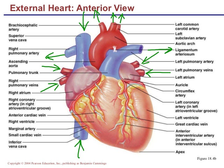
External structure of the heart with labels. Internal Structure of the Heart | Contemporary Health Issues Internal Structure of the Heart. Recall that the heart's contraction cycle follows a dual pattern of circulation—the pulmonary (lungs)and systemic (body) circuits—because of the pairs of chambers that pump blood into the circulation. In order to develop a more precise understanding of cardiac function, it is first necessary to explore the ... Heart Anatomy | Anatomy and Physiology II - Lumen Learning Describe the location and position of the heart within the body cavity; Describe the internal and external anatomy of the heart; Identify the tissue layers of the heart; Relate the structure of the heart to its function as a pump; Compare systemic circulation to pulmonary circulation; Identify the veins and arteries of the coronary circulation ... Solved Art-Labeling Activity: Overview of the external - Chegg art-labeling activity: overview of the external anatomy of the heart anterior view res great cardiac vein aortic arch right coronary artery left coronary artery left pulmonary veins ascending aorta left pulmonary artery anterior interventricular artery superior vena cava pulmonary trunk auricle of left atrium circumflex artery auricle of right … Structure Of The Heart | A-Level Biology Revision Notes The heart is a hollow muscular organ that lies in the middle of the chest cavity. It is enclosed in the pericardium, which protects the heart and facilitates its pumping action. The heart is divided into four chambers: The two atria (auricles): these are the upper two chambers. They have thin walls which receive blood from veins.
Label the heart — Science Learning Hub Label the heart Interactive Add to collection In this interactive, you can label parts of the human heart. Drag and drop the text labels onto the boxes next to the diagram. Selecting or hovering over a box will highlight each area in the diagram. Aorta Vena cava Right ventricle Semilunar valve Left atrium Left ventricle Right atrium Pulmonary vein 2. External features of the heart - SlideShare 2. THE HEART • The heart is a hollow muscular organ that is pyramidal in shape • It lies within the pericardium in the middle mediastinum • It is connected at its base to the great blood vessels. 3. General features of the heart • The heart has; • an apex and base • 2 surfaces; • Sternocostal surface • Diaphragmatic surface ... The Tenors - Wikipedia The Tenors (formerly known as The Canadian Tenors) are a vocal group consisting of Victor Micallef, Clifton Murray, Alberto Urso, and Mark Masri.They perform operatic pop music that is a mixture of classical and pop, featuring songs such as "The Prayer", Panis angelicus, and Leonard Cohen's Hallelujah. Heart Anatomy: size, location, coverings and layers : Anatomy & Physiology Heart Anatomy. The heart is around the size of a fist and weighs between 250-350 grams (less than a pound). Enclosed within the mediastinum, the medial cavity of the thorax, the heart extends obliquely from the second rib to the fifth intercostal space. It rests on the superior surface of the diaphragm, lies posterior to the sternum and ...
Dapagliflozin - Wikipedia Medical uses. Dapagliflozin is used along with diet, exercise and usually with other glucose lowering medications, to improve glycaemic control in adults with type 2 diabetes and to reduce the risk of hospitalisation for heart failure among adults with type 2 diabetes and known cardiovascular disease or other cardiovascular risk factors (including high blood pressure, high cholesterol and ... Structure of the Heart | SEER Training - National Cancer Institute The human heart is a four-chambered muscular organ, shaped and sized roughly like a man's closed fist with two-thirds of the mass to the left of midline. The heart is enclosed in a pericardial sac that is lined with the parietal layers of a serous membrane. The visceral layer of the serous membrane forms the epicardium. Layers of the Heart Wall A Labeled Diagram of the Human Heart You Really Need to See The human heart, comprises four chambers: right atrium, left atrium, right ventricle and left ventricle. The two upper chambers are called the left and the right atria, and the two lower chambers are known as the left and the right ventricles. The two atria and ventricles are separated from each other by a muscle wall called 'septum'. Diagram of Human Heart and Blood Circulation in It The outermost layer of your heart wall is called the epicardium, which is basically a very thin layer of serous membrane. The membrane provides lubrication and protection to the outer side of your heart, as you can see in heart diagram labeled. Myocardium Right beneath epicardium is another relatively thicker layer called myocardium.
Ap2 chap18heartclass
Heart Anatomy: Labeled Diagram, Structures, Blood Flow ... - EZmed Let's begin with the chambers of the heart. There are 4 chambers, labeled 1-4 on the diagram below. To help simplify things, we can convert the heart into a square. We will then divide that square into 4 different boxes which will represent the 4 chambers of the heart.

Heart and blood vessels - online presentation
Chapter 22 Heart Flashcards | Quizlet Label the coronary arteries in an anterior view of the heart. Label the order that blood flows through in the heart, using the arrows as guides. Label the components of the heart wall. Label the components of the heart as seen from a posterior view. Label the major coronary veins. Label the components of the conduction system.

Medical Encyclopedia - Structure: Structure of the Heart - Aviva | THE HEART | Pinterest | Heart ...
Self-esteem - Wikipedia Moral judgment stages: Individuals describe their real, ideal, and dreaded selves with stereotypical labels, such as "nice" or "bad". Individuals describe their ideal and real selves in terms of disposition for actions or as behavioral habits. The dreaded self is often described as being unsuccessful or as having bad habits.
Heart Anatomy Cross Section Diagram Stock Vector 157286378 - Shutterstock
Heart - External Features - Anatomy QA Location of heart: Heart lies in the middle mediastinum. 1/3rd of the heart lies to the right and 2/3rd to the left of the midline. It lies opposite to T5 - T8 vertebrae in supine position & T6 - T9 vertebrae in erect position. Dimensions of heart: Base to apex-12cm; Transversely- 8-9cm; Anteroposteriorly- 6cm.

Aqua Fanatic: 07/01/2011 - 08/01/2011
Heart Anatomy Labeling Game - PurposeGames.com This is an online quiz called Heart Anatomy Labeling Game. There is a printable worksheet available for download here so you can take the quiz with pen and paper. Your Skills & Rank. Total Points. 0. Get started! Today's Rank--0. Today 's Points. One of us! Game Points. 19. You need to get 100% to score the 19 points available.

The Science Scoop: Heart Diagram
Heart Diagram with Labels and Detailed Explanation - BYJUS Diagram of Heart. The human heart is the most crucial organ of the human body. It pumps blood from the heart to different parts of the body and back to the heart. The most common heart attack symptoms or warning signs are chest pain, breathlessness, nausea, sweating etc. The diagram of heart is beneficial for Class 10 and 12 and is frequently ...
Human Heart Drawing Images at GetDrawings | Free download
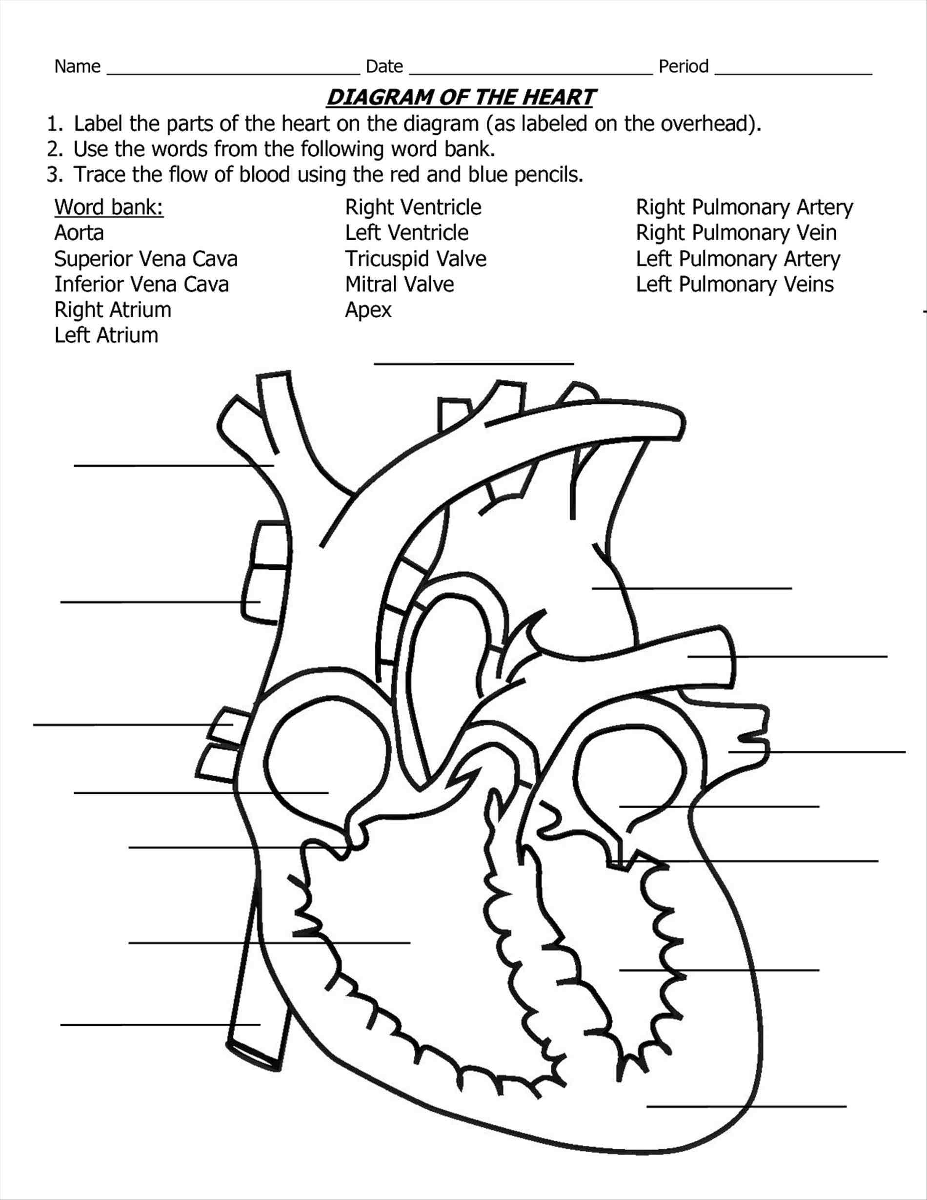
Label the Heart - The Biology Corner Shows a picture of a heart with letters and blanks for practice with labeling the parts of the heart and tracing the flow of blood within the heart.

32 Label The Anterior View Of The Human Heart - Labels Design Ideas 2020
Diagram of the human heart royalty-free images - Shutterstock 14,791 diagram of the human heart stock photos, vectors, and illustrations are available royalty-free. See diagram of the human heart stock video clips. Image type.

External Structure Of Human Heart Anatomy | MedicineBTG.com
The Anatomy of the Heart, Its Structures, and Functions - ThoughtCo The heart is the organ that helps supply blood and oxygen to all parts of the body. It is divided by a partition (or septum) into two halves. The halves are, in turn, divided into four chambers. The heart is situated within the chest cavity and surrounded by a fluid-filled sac called the pericardium. This amazing muscle produces electrical ...

Aqua Fanatic: Crayfish Anatomy
How Strategy Shapes Structure - Harvard Business Review See Industrial Market Structure and Economic Performance, F. M. Sherer (Chicago: Rand McNally, 1970). 2. See Blue Ocean Strategy , W. Chan Kim and Renée Mauborgne (Harvard Business Press, 2005).

the unlabelled structure of heart - Google Search | Heart diagram, Parts of the heart, Nursing ...
Correctly Label The Following External Anatomy Of The Anterior Heart ... The external anatomy of the human heart consists of the four chambers that form the apex of the heart. Each chamber has an apex that corresponds to a box. There are two boxes on each side of the heart: the atria and the ventricles. The left atrium is a branching organ. The pulmonary trunk contains the aorta and pulmonary veins.
😀 Drag the labels to identify structural components of the heart. Label the heart — Science ...
Chapter 19: The Heart Flashcards | Quizlet •Allows heart to beat without friction, gives it room to expand and resists excessive expansion •Parietal pericardium-tough outer, fibrous layer of connective tissue-inner serous layer •Visceral pericardium (a.k.a. epicardium of heart wall)-serous lining of sac turns inward at base of heart to cover the heart surface
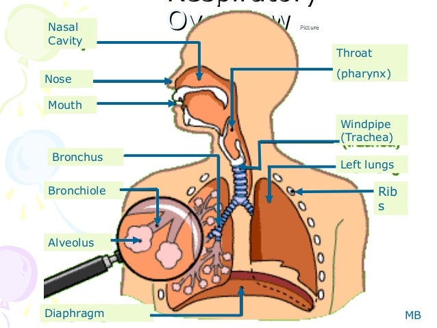
Human respiration
Layers of the heart: Epicardium, myocardium, endocardium - Kenhub The endocardium is the innermost layer of the heart. It lines the inner surfaces of the heart chambers, including the heart valves. The endocardium has two layers. The inner layer lines the heart chambers and is made of endothelial cells.
32 Label The Diagram Of The Heart - Labels Database 2020
Solved Help Label the external anatomy on this posterior - Chegg Question: Help Label the external anatomy on this posterior view of a mammalian heart by clicking and dragging the labels to the correct location Coronary sinus Apex of heart Lert atrium Posterior interventricular branch of LCA Left pulmonary artery Left ventricle Left pulmonary veins Aortic arch This problem has been solved! See the answer

The Heart | S-cool, the revision website
Human Heart Diagram Labeled | Science Trends The endocardium is the inner portion of the outer wall, and the endocardium is what contacts the blood in the heart. The heart's atrioventricular valves are structures that join the atria and ventricles of the heart together. This group of valves is comprised of the tricuspid valve and the mitral valve.

Cardiovascular System - Anatomy And Physiology
Human Heart - Diagram and Anatomy of the Heart - Innerbody Because the heart points to the left, about 2/3 of the heart's mass is found on the left side of the body and the other 1/3 is on the right. Anatomy of the Heart Pericardium. The heart sits within a fluid-filled cavity called the pericardial cavity. The walls and lining of the pericardial cavity are a special membrane known as the pericardium.
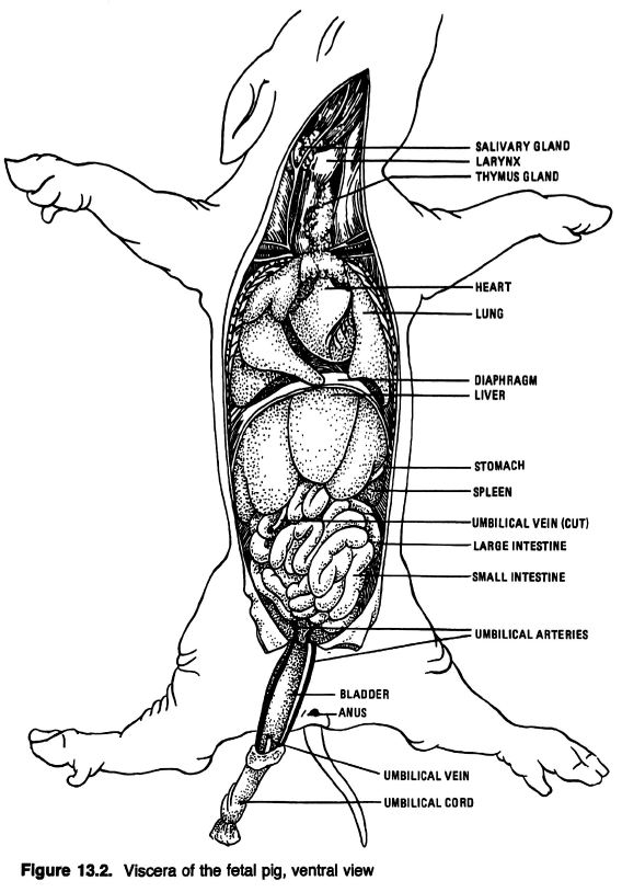
Anatomical Drawings of a Fetal Pig
Heart Anatomy: Heart Dissection - University of Washington The letters indicated in the text refer to the labels on the picture. The anterior surface of the heart is characterized by the presence of the large arteries leaving the base of the heart, the pulmonary trunk (H) and the aorta (G). The pulmonary trunk is the vessel that divides to give rise to the two pulmonary arteries going to each lung.

Biology 156: April 2012
External anterior heart labeling Quiz - purposegames.com About this Quiz This is an online quiz called External anterior heart labeling There is a printable worksheet available for download here so you can take the quiz with pen and paper. Your Skills & Rank Total Points 0 Get started! Today's Rank -- 0 Today 's Points One of us! Game Points 27 You need to get 100% to score the 27 points available
Post a Comment for "44 external structure of the heart with labels"