38 sheep heart dissection pictures with labels
Sheep Cardiovascular (SCA_001) - Bio Medical Images Ventral aspect of sheep heart before dissectionLabels included below. Price includes a one time usage. You will get immediate access to the file and for 30 days after purchase.LABELS: 1. Right auricle2. Pulmonary trunk3. Fat (adipose tissue)4. Left auricle5. Anterior sulcus6. Right ventricle7. Left ventricle8. Apex of the heart sheep heart dissection.docx - Picture 1 External View Dorsal side(label ... View sheep heart dissection.docx from ANATOMY & 2513 at Pearl River Community College. Picture 1 - External View Dorsal side (label dorsal side). Picture 2- External View Ventral side (label ventral Study Resources Main Menu by School by Literature Title by Subject Textbook SolutionsExpert TutorsEarn Main Menu Earn Free Access Upload Documents
heart anatomy diagram unlabeled The Heart Diagrams Labeled and Unlabeled we have 9 Pics about The Heart Diagrams Labeled and Unlabeled like Heart Diagram - 15+ Free Printable Word, Excel, EPS, PSD Template, The Heart Diagrams Labeled and Unlabeled and also Respiratory System Worksheet - WikiEducator. Read more: The Heart Diagrams Labeled And Unlabeled

Sheep heart dissection pictures with labels
Sheep Heart - San Diego Mesa College SHEEP HEARTS. Click on a photo for a larger view of the model. Click on Label for the labeled model. Back to Dissected Specimen Page. Anterior view: Arteries: Posterior view: Label: Label: Label: Venae cavae: Medial view: Valves-Right: Label: Label: Label: Tricuspid valve: Longitudinal cut: Chambers cut: Label: The Ultimate Heart Model & Sheep Heart Practice Quiz! - ProProfs Take this practice quiz that covers information related to the sheep heart & the heart model. It is intended for use as a supplemental study aid. As is the case in the lab practical, each correct answer counts. So, make sure you learn from the feedback. Questions and Answers 1. 1. Name the structure- be specific. 2. 2. Name the vessel- be specific. Sheep Brain Dissection with Labeled Images Sheep Brain Dissection with Labeled Images Sheep Brain Dissection 1. The sheep brain is enclosed in a tough outer covering called the dura mater. You can still see some structures on the brain before you remove the dura mater. Take special note of the pituitary gland and the optic chiasma.
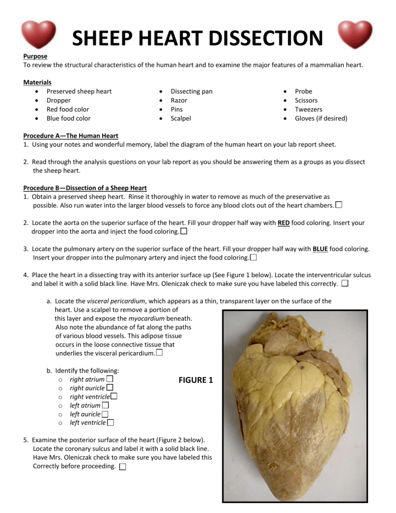
Sheep heart dissection pictures with labels. ANATOMY- Sheep HEART DISSECTION - ANJA'S AICE ANATOMY- Sheep HEART DISSECTION - ANJA'S AICE Sheep Heart Dissection Grace Boshart and Anja Stichter Lab Report 1. Purpose: To get a better understanding of the mammalian heart. We were able to make connections between what we had learned about the structure and function of the heart with what we could observe on a real heart. 2. Sheep Heart Images - San Diego Mesa College SHEEP HEART IMAGES. Sheep Heart Unlabeled. Sheep Heart Leader-lined. Sheep Heart Labeled. San Diego Mesa College 7250 Mesa College Drive San Diego, CA 92111-4998 Student Support San Diego Community College District San Diego City College San Diego Mesa College San Diego Miramar College PDF Sheep Heart Dissection Lab - Mr. E. Science c. Use the scalpel and slice the heart, almost in half, as shown in figure 3. Slice thru the middle of the right atrium across the top of the heart across to the left atrium. Continue the cut down the left atrium thru the left ventricle to the apex of the heart. Gentle pull open the heart exposing the internal structure. Continue slicing through Title: Sheep Heart Dissection - SlideServe Title: Sheep Heart Dissection Objective: -To Practice Dissection -To discover and explore anatomical parts of the heart Materials: Sheep Heart, Dissection Tray, Manual (Pictures), Scalpel, Gloves Procedure: • Obtain heart and dissection tools. • Find Ventral surface, sketch and label items in lab book • Make longitudinal section (cut), observe section, bisected heart labeling items in ...
Cardiovascular Cat Dissection Labeled - Virtual Anatomy Lab Cardiovascular Models Unlabeled. Cardiovascular Sheep Heart Dissect-L. Cardiovascular Sheep Heart Disect-U. Cardiovascular Cat Dissection Labeled. Cardiovascular Cat Dissection Unlabeled. Cardiovascular Rabbit Dissection-L. Cardiovascular Rabbit Dissection -U. Respiratory. Respiratory Histology Labeled. Sheep Heart Dissection - Josh Li Anatomy and Physiology Voles and humans also have canines and molars, similar to humans. A major key difference is the size of the skull. The average human skull ranges from 6-7 inches while the skull of a vole is only 25mm. Humans have teeth all across our whole mouths while a vole as sharp teeth in the upper and lower parts of the jaw. Sheep Heart Dissection Projects | Photos, videos, logos, illustrations ... Behance is the world's largest creative network for showcasing and discovering creative work Heart Dissection : 8 Steps (with Pictures) - Instructables There our other dissection photos out there, but I wanted to make a clear walkthrough for teachers and students who are doing it. What: Heart Dissection. Concepts: biology, anatomy, pumps. Time: ~45-60 minutes. Cost: ~$1.50 per heart. Materials: One heart (pig, cow, or sheep from the butcher as in tact as possible) 4 x Dowels (or pencils)
Heart Dissection | Carolina.com Heart Dissection | Carolina.com Login or Register 800.334.5551 My Account Login or register now to maximize your savings and access profile information, order history, tracking, shopping lists, and more. Login Create an Account Service & Support Contact Us Our Customer Service team is available from 8am to 6:30pm, ET, Monday through Friday. Sheep Heart Dissection Lab for High School Science | HST Dissection: Internal Anatomy 1. Insert your dissecting scissors or scalpel Click for full-size pdf into the superior vena cava and make an incision down through the wall of the right atrium and ventricle, as shown by the dotted line in the external heart picture. Pull the two sides apart and look for three flaps of the membrane. Sheep Heart Dissection Teaching Resources | Teachers Pay Teachers Get the most out of your sheep heart dissection. Complete student background information is included with labeled images for identifying internal and external sheep heart anatomical structures. No prior knowledge of heart anatomy is required. Students learn both internal and external anatomy of the heart, the flow of blood through the heart, a PDF Virtual Sheep Heart Dissection Lab - MRS. MERRITT'S ANATOMY CLASS Johnson's Sheep Heart Dissection" posted by stacyelambert Purpose: To examine the major features of a mammalian heart. ... The sheet with the pictures are in class. You will find it where the YODA poster is on the ... Label the interior structures of the heart in Figure 3: Pick up the document from class; the pics were too large to upload. ...
PDF Name: Dissection 101: Sheep Heart - PBS The sheep heart is mammalian, having four chambers like the human heart, which includes two atria and two ventricles. The blood flow through the sheep heart is like that of the human heart, in which the blood is pumped from the right side of the heart to the lungs and then from the left side of the heart to the body. Orientation of the heart: •
heart dissection-lab report.doc - Name: Date: Period: Sheep Heart ... - There should only be 6 total pictures.- The pictures should be in the order below with only the stated labels. Delete the above information in your completed lab report Picture #1 - (ventral view): coronary arteries/veins, base, apex, myocardium, adipose, visceral pericardium
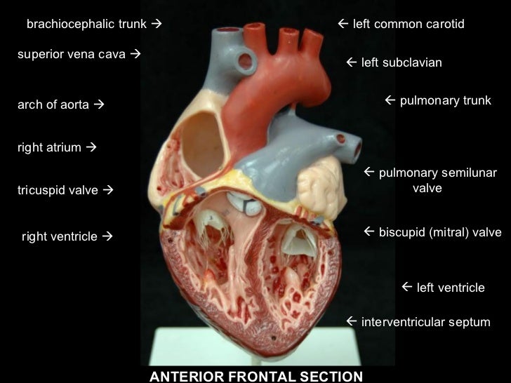
Heart Dissection Photos - Valdosta State University Heart Dissection Photos. Click on the thumbnails to see the large labeled images. Click the back button on your browser to return to this list. Pericardium. Anterior surface. Posterior surface. Left ventricle open. Left ventricle with probes. Arteries vs. veins. Vena cavae. Right ventricle open. Semilunar valves. Coronary sinus. Bicuspid valves
Sheep brain dissection and label - slideshare.net Sheep brain-dissection and label By Leslie Young Section 63 2. Sheep brain-dissected and shown olfactory bulb and optic nerve. The olfactory bulb is a structure of the vertebrate forebrain involved in olfaction, the perception of odors The optic nerve, also known as cranial nerve 2, transmits visual information from the retina to the brain. 3.
PDF Sheep Heart Dissection Lab - Home Science Tools Sheep Heart Dissection Kit Here Background Sheep have a four-chambered heart, just like humans. ... Label the Parts of a Sheep Heart ... label the left side (.pdf) See our other free dissection guides with photos and printable PDFs. Click here. Sheep Heart issection 4/6. Sheep Heart issection 5/6. Sheep Heart issection 6/6. Created Date: 4/23 ...
Virtual Sheep Heart Dissection Lab Answer Key Read Free Virtual Sheep Heart Dissection Lab Answer Key draw, label, apply clinical content, and think critically, Wood, Laboratory Manual for Anatomy & Physiology featuring Martini Art , Main Version, Fifth Edition offers a comprehensive approach to the two-semester A&P laboratory course. The stunning, full-color illustrations are adapted from
How would you label the structures (both external and internal) of a dissected pig's heart? - Quora
Heart Dissection High Resolution Stock Photography and Images - Alamy Find the perfect heart dissection stock photo. Huge collection, amazing choice, 100+ million high quality, affordable RF and RM images. ... Medical students doing sheep heart dissection in the lab class Close up view of a partially dissected cow heart as used in a UK high school demonstration. ... Search Results for Heart Dissection Stock ...
PDF Sheep Heart Dissection - Mrs. Moretz's Science Site Sheep Heart Dissection Procedure (Day 2) - you will be cutting the heart open today! a. Review the outer part of the heart and make sure you know where these structures are: left and right ventricle, left and right atrium, pulmonary artery, pulmonary veins, aorta, coronary artery, apex, superior and inferior vena cava. b. Locate the pulmonary ...
Heart Diagram Labeled Igcse : The Human Eye Edexcel Igcse Biology ...
Dissection of the Sheep Heart - Houston Community College Dissection of the Sheep Heart 1 Dissection of the Sheep Heart and Human Heart INTRODUCTION The heart is a cone-shaped muscular organ, about the size of your fist. It is located in the mediastinum region (central region of the thoracic cavity), between the lungs, and behind the sternum. The heart is a hollow organ, containing 4 chambers.
Sheep Heart Dissection - The Biology Corner Sheep Heart Dissection External Anatomy 1. Identify the right and left sides of the heart. Look closely and on one side you will see a diagonal line of blood vessels that divide the heart, this line is called the interventricular sulcus. The half that includes all of the apex (pointed end) of the heart is the left side. 2.
Sheep Brain Dissection with Labeled Images Sheep Brain Dissection with Labeled Images Sheep Brain Dissection 1. The sheep brain is enclosed in a tough outer covering called the dura mater. You can still see some structures on the brain before you remove the dura mater. Take special note of the pituitary gland and the optic chiasma.
The Ultimate Heart Model & Sheep Heart Practice Quiz! - ProProfs Take this practice quiz that covers information related to the sheep heart & the heart model. It is intended for use as a supplemental study aid. As is the case in the lab practical, each correct answer counts. So, make sure you learn from the feedback. Questions and Answers 1. 1. Name the structure- be specific. 2. 2. Name the vessel- be specific.

Labeled Sheep Heart Picture #1 | Edu Circulatory System | Heart pictures, Circulatory system ...
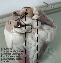
Sheep Heart - San Diego Mesa College SHEEP HEARTS. Click on a photo for a larger view of the model. Click on Label for the labeled model. Back to Dissected Specimen Page. Anterior view: Arteries: Posterior view: Label: Label: Label: Venae cavae: Medial view: Valves-Right: Label: Label: Label: Tricuspid valve: Longitudinal cut: Chambers cut: Label:


Post a Comment for "38 sheep heart dissection pictures with labels"