40 brain mri with labels
Brain MRI: How to read MRI brain scan | Kenhub MRI is the most sensitive imaging method when it comes to examining the structure of the brain and spinal cord. It works by exciting the tissue hydrogen protons, which in turn emit electromagnetic signals back to the MRI machine. The MRI machine detects their intensity and translates it into a gray-scale MRI image. parts of the brain labeled Mri Brain, Mri, Radiology Imaging . mri brain anatomy sagittal labeled mrimaster cross sectional coronal radiology mr randal versus head labels imaging koene gradual identity personal. Untitled Document [bio.sunyorange.edu] bio.sunyorange.edu. brain cerebellum pig dissection anatomy development bio human adolescence childhood
brain and parts labeled brain and parts labeled. Mri eye anatomy muscles scan sciencephoto. Diencephalon and brain stem: unit 4, group 3. Brain sagittal anatomy human section labeled cat robotspacebrain cut diagram parts physiology medical mid function mri structure labels drawing fig. brain and parts labeled.

Brain mri with labels
Brain: Atlas of human anatomy with MRI - e-Anatomy - IMAIOS MRI Atlas of the Brain. This page presents a comprehensive series of labeled axial, sagittal and coronal images from a normal human brain magnetic resonance imaging exam. This MRI brain cross-sectional anatomy tool serves as a reference atlas to guide radiologists and researchers in the accurate identification of the brain structures. Learning-based 3T brain MRI segmentation with guidance from 7T MRI labeling Learning-based 3T brain MRI segmentation with guidance from 7T MRI labeling Med Phys. 2016 ... Therefore, they are not ideal for providing good ground truth label data for training learning-based methods. Recent advances in ultrahigh field 7T imaging make it possible to acquire images with excellent intensity contrast and signal-to-noise ratio. ... CaseStacks.com - MRI Brain Anatomy Labeled scrollable brain MRI covering anatomy with a level of detail appropriate for medical students. Show/Hide Labels. MRI Brain Anatomy. Back to Anatomy overview. Facebook; Twitter; ... Labelled radiographs and CT/MRI series teaching anatomy with a level of detail appropriate for medical students and junior residents. Pelvis. Pelvic MRI anatomy
Brain mri with labels. Brain lobes - annotated MRI | Radiology Case | Radiopaedia.org 100 public playlists include this case. CT KUB by kazi moin uddin. Learning-SectionalAnatomy by Dr Payam Riahi. Anatomy - Neuro, Head & Neck by Dr Yair Glick . Barin by Dr nermin Nermin Hassan Aboyoussef. 2022 4 by Richard Hodgson. Brain - Anatomy by Dwayne Ian Reading. Anatomy by Mariangela Alvarado Molinaro. Neuro Essentials by Duaa Kanan. 101 Labeled Brain Images and a Consistent Human Cortical Labeling ... Labeled anatomical subdivisions of the brain enable one to quantify and report brain imaging data within brain regions, which is routinely done for functional, diffusion, and structural magnetic resonance images (f/d/MRI) and positron emission tomography data. GitHub - yunshiuan/label4MRI: Label the brain MNI coordinate by AAL/BA ... MRI-labeling: label human brain MRI image by AAL/BA system Under the R program environment,input an MNI coordinate, output the corresponding AAL(Automated Anatomical Labeling) and BA (Brodmann area) brain region name. More importantly, if the coordinate does not match a brain region defined by AAL/BA (e.g., white matter), the package help find the closest brain region with the corresponding ... MRI head axial T2 - labeling questions - Radiopaedia The labeled structures are (excluding the correct side): cervical spinal cord posterior arch of C1 odontoid process (peg or dens) of C2 parotid gland intradural segment (V4) of dominant vertebral artery cisterna magna intradural segment (V4) of non-dominant vertebral artery cerebellar tonsil occipital condyle medulla oblongata
MR Image Classification for Brain Tumor Texture Based on Pseudo-Label ... MR Image Classification for Brain Tumor Texture Based on Pseudo-Label Learning and Optimized Feature Extraction Comput Math Methods Med. 2022 Apr 4;2022:7746991. doi: 10.1155/2022/7746991. ... First, for the small sample of pituitary tumor MRI image data, the T1 and T2 sequence data are uneven or missing; we used the CycleGAN model to perform ... MRI Brain Atlas This web app Atlas is intended for veterinary students and radiologists seeking quick access to canine brain anatomy through a mobile device. Via a toggle button, either MRI images or approximately comparable Brain Transection images may be viewed with or without labels. Navigation & Labels. Cross-sectional anatomy of the brain - e-Anatomy - IMAIOS We created a brain atlas that is an interactive tool for studying the conventional anatomy of the normal brain based on a magnetic resonance imaging exam of the axial brain. Anatomical structures and specific areas are visible as interactive labeled images. Cross sectional anatomy: MRI of the brain. An MRI was performed on a healthy subject ... Atlas of BRAIN MRI - W-Radiology Brain magnetic resonance imaging (MRI) is a common medical imaging method that allows clinicians to examine the brain's anatomy (1). It uses a magnetic field and radio waves to produce detailed images of the brain and the brainstem to detect various conditions (2).
Labeled MRI Brain Scans - Neuromorphometrics We can also label scans that you provide and we are very interested in labeling white matter anatomy as seen in diffusion-weighted MRI scans. If you want an aggregate version of our data, we can provide it as a probabilistic atlas. The cost to label a single scan is $2449 (USD). MRI anatomy | free MRI axial brain anatomy MRI anatomy | free MRI axial brain anatomy This MRI brain cross sectional anatomy tool is absolutely free to use. Use the mouse scroll wheel to move the images up and down alternatively use the tiny arrows (>>) on both side of the image to move the images. A normative spatiotemporal MRI atlas of the fetal brain for automatic ... Step 2: The segmented neonatal atlases were used to generate initial labels on the spatiotemporal fetal brain MRI atlas at higher GAs (35-37 weeks) through multiatlas segmentation using probabilistic label fusion 65. Step 3: Fetal brain MRI labels were manually defined and propagated in iterations from the higher GAs to the lower GAs. Deep learning from MRI-derived labels enables automatic brain tissue ... Deep learning from MRI-derived labels enables automatic brain tissue classification on human brain CT Neuroimage. 2021 Dec 1;244:118606. doi: 10.1016/j ... Our proposed model predicted brain tissue classes accurately from unseen CT images (Dice coefficients of 0.79, 0.82, 0.75, 0.93 and 0.98 for GM, WM, CSF, brain volume and ICV, respectively). ...
normal mri top brain - Google Search | ANATOMY | Pinterest | Tops and Search
Labeled imaging anatomy cases | Radiology Reference Article ... This article lists a series of labeled imaging anatomy cases by body region and modality. Brain CT head: non-contrast axial CT head: non-contrast coronal CT head: non-contrast sagittal CT head: angiogram axial CT head: angiogram coronal CT...

Normal Brain CT MRI Imaging ( In Arabic ) Prof Dr Mamdouh Mahfouz - YouTube
brain anatomy | MRI coronal brain anatomy | free MRI cross sectional ... This MRI brain coronal cross sectional anatomy tool is absolutely free to use. Use the mouse scroll wheel to move the images up and down alternatively use the tiny arrows (>>) on both side of the image to move the images.>>) on both side of the image to move the images.

Making My Theology Work: An Introduction - ParkingSpace23ParkingSpace23
NITRC: Manually Labeled MRI Brain Scan Database: Tool/Resource Info This is a continuously growing and improving database of high-quality neuroanatomically labeled MRI brain scans, created not by an algorithm, but by neuroanatomical experts. All results are checked and corrected. Regions of interest include the usual sub-cortical structures (thalamus, caudate, putamen, hippocampus, etc), along with ventricles ...

Images of Cadaver and Human Brain of 7.0 T MRI In Vivo | Radiology Key
Researchers automate brain MRI image labeling, more than 100,000 exams ... Researchers have automated brain MRI image labeling, needed to teach machine learning image recognition models, by deriving important labels from radiology reports and accurately assigning them to...

MRI BLOG: Pitfalls of Diffusion Weighted Imaging
CaseStacks.com - MRI Brain Anatomy Labeled scrollable brain MRI covering anatomy with a level of detail appropriate for medical students. Show/Hide Labels. MRI Brain Anatomy. Back to Anatomy overview. Facebook; Twitter; ... Labelled radiographs and CT/MRI series teaching anatomy with a level of detail appropriate for medical students and junior residents. Pelvis. Pelvic MRI anatomy
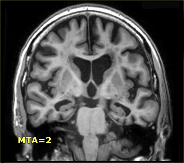
The Radiology Assistant : Brain - Dementia: Role of MRI
Learning-based 3T brain MRI segmentation with guidance from 7T MRI labeling Learning-based 3T brain MRI segmentation with guidance from 7T MRI labeling Med Phys. 2016 ... Therefore, they are not ideal for providing good ground truth label data for training learning-based methods. Recent advances in ultrahigh field 7T imaging make it possible to acquire images with excellent intensity contrast and signal-to-noise ratio. ...

Bl4ck D4ys: March 2011
Brain: Atlas of human anatomy with MRI - e-Anatomy - IMAIOS MRI Atlas of the Brain. This page presents a comprehensive series of labeled axial, sagittal and coronal images from a normal human brain magnetic resonance imaging exam. This MRI brain cross-sectional anatomy tool serves as a reference atlas to guide radiologists and researchers in the accurate identification of the brain structures.

Brain, MRI - Stock Image - C036/6075 - Science Photo Library

MRI scan of brain - Stock Image - P332/0482 - Science Photo Library
Test 1 | Radiology Key

How large is your brain? MRI - YouTube

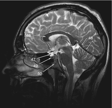
The Radiology Assistant : Brain Anatomy | Brain anatomy, Radiology imaging, Mri brain

MRI BLOG: Pitfalls of Diffusion Weighted Imaging

Multiple sclerosis | Image | Radiopaedia.org

Brain MRI Quiz

Dr Balaji Anvekar FRCR: Superior cerebellar territory infarct
Psych 104 Chapter 3 Lecture 2
Post a Comment for "40 brain mri with labels"